MODULE 1 - SECTION 4 - CEREBROSPINAL FLUID, VENTRICLES and 3 SPACES
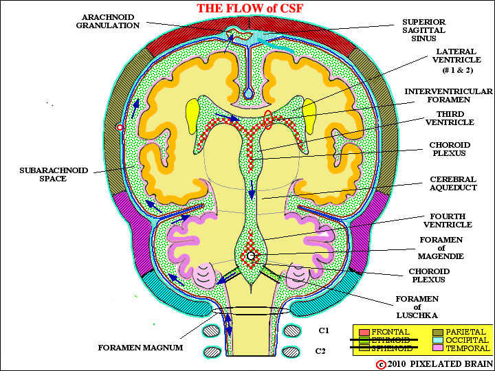
In section 2 (Brain Development) we learned that the brain is really a tube-like structure, formed when the neural plate rolls up and the edges meet to enclose a central cavity. The cavity becomes the ventricular system of the adult brain. In certain regions the edges of the plate fail to meet, and vascular tissue bridges the gap. This tissue is named the choroid plexus and it produces the cerebrospinal fluid (CSF) that fills the ventricular system and escapes into the space between the brain's surface and the skull.
Fenestrated capillaries within choroid tissue allow for free passage of water and solutes but that tight junctions between choroid epithelial cells form a blood-CSF barrier. While lipid soluble substance may easily pass through this barrier, most other materials must be conveyed from blood to CSF by active transport through cells of the choroid epithelium. The result is that the chemical composition of the two fluids differ; for example, CSF has higher levels of chloride, magnesium and sodium, compared with plasma. CSF is colorless, low in protein and contains very few, if any, white blood cells.
The volume of CSF in the adult is about 120mL and roughly 500mL of the fluid is produced every day, so that about 4 turnovers occur in 24 hour. Production continues even if there is a blockage of flow, somewhere along the way to the arachnoid granulations, but the result depends on the age of the subject and the site of the obstruction.
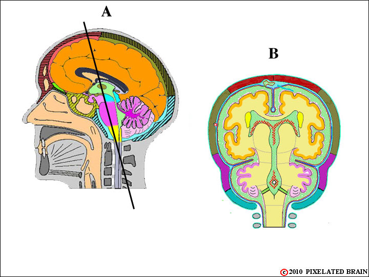
Some of these details are illustrated in the views below.
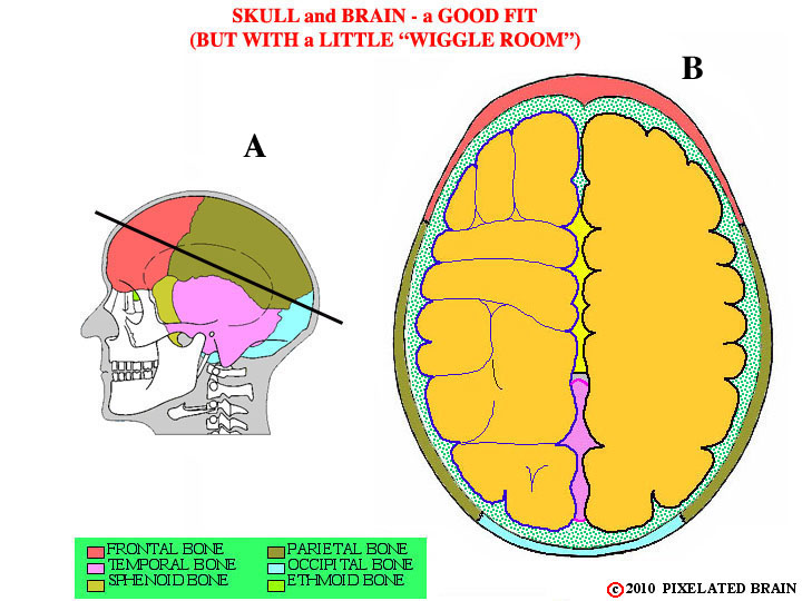
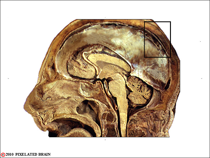
Certainly, the most important function of the skull is to protect the fragile brain, lying within the cranial cavity. As this figure indicates, the skull is well-designed for the job. If we cut through the calvarium in the plane shown in A and look down from above we get view B.
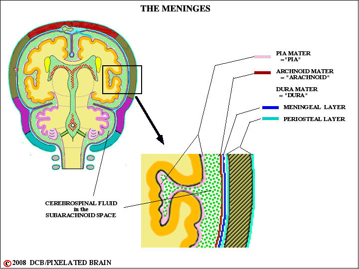
The contour of the brain's surface matches that of the cranial cavity rather well, but there is a small space between the two. This narrow gap, shown by green stippling in the figure, is called the subarachnoid space. It is filled with cerebrospinal fluid (CSF) and also contains three membranes known collectively as the meninges. The brain is, in a sense, floating in CSF which protects it from "bumping into" the skull during sudden movements of the head.
From a clinical point of view, the anatomy of this region is extremely important and we want to look at it in detail. The best way to start is to consider CSF - where does this fluid come from, how does it get into the subarachnoid space, where is it going, and how does all this relate to the meninges?
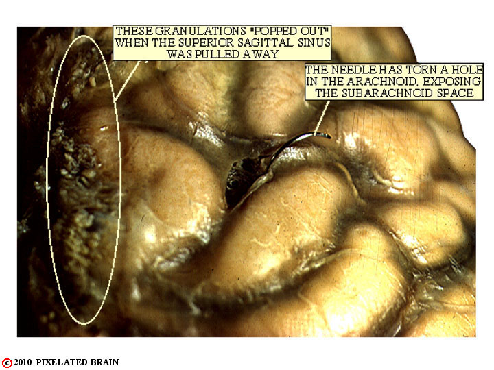
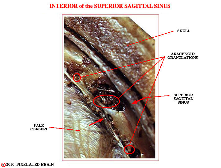
Granulations are easily seen in gross specimens. If the dura is carefully removed from the surface of the brain, they tend to pop out of the superior sagittal sinus and be exposed to view, as shown in this view.
THE MENINGEAL SPACES AND INTRACRANIAL BLEEDIN:
THE THREE SPACES
The meninges, by their very existence, define three clinically important spaces.
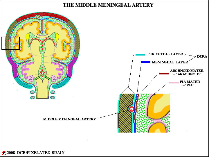
1) The epidural space is a potential one because the dura normally lies in direct contact with the inner surface of the skull. However, when skull fractures occur and the fracture line crosses the valley which the middle meningeal artery erodes in the skull then there is a good chance that the vessel will be torn. If this happens, the position of the artery is such (see figure ) that blood will force its way between the dura and the skull, creating an epidural hematoma. Because the dura is adherent to the skull, the accumulating mass assume a characteristic "lens" shape as blood dissects the dura away from the skull. Take a look at the You Tube video
2) The Subdural Space
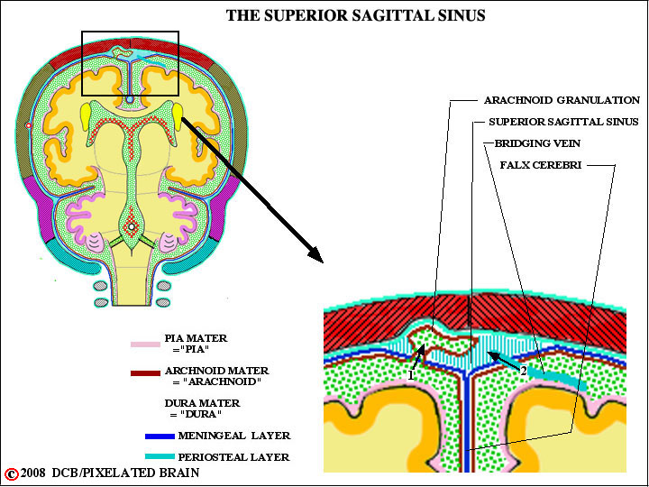
The subdural space lies between the dura and the arachnoid and is also only a potential one, since the pressure of CSF normally presses the arachnoid against the dura. However if, in an accident, the skull strikes a hard surface (the dashboard of a car), the brain may shift position slightly within the cranial cavity and as a result the bridging veins, passing from the surface of the hemisphere into the superior sagittal sinus (see figure ) may be torn. These veins pass through the subdural space, and that's where the blood accumulates. Since the the dura and arachnoid easily separate, the blood in this case spreads easily over the surface of the hemisphere (see Blumenfeld pg 166 for a typical lesion). Take a look at the You Tube video.
3) The Subarachnoid Space
The subarachnoid space is, of course, a real one, normally filled with CSF, but the arteries supplying the brain run in this space. When one ruptures, blood escapes into the space and spreads diffusely.