MODULE 2 - SECTION 3 - VENTRAL and SAGITTAL VIEWS of the BRAINSTEM
BRAINSTEM VENTRAL VIEW
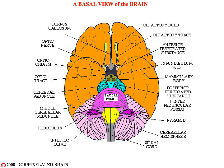
While the dorsal surface of the brainstem is normally hidden from view by overlying structures (cerebral hemispheres, cerebellum) the ventral (or basal) surface of the brainstem is exposed in views of the whole brain, like this one.
Here, as elsewhere in this module, we include the diencephalon in our discussion of brainstem anatomy, even though - strictly speaking - it is not part of the brainstem. Also, we may at times refer to the diencephalon as the thalamus, even though this is not quite right either.
This is a good point to pause and look at some excellent views taken from The Digital Anatomist:
Diganat_1f - view of gross brain, very similar to this drawing of one
Diganat_5c - ventral view of a dissected brain
Diganat_5e - oblique view of a dissected brain
Diganat_5j - best shot of all - don't miss it!
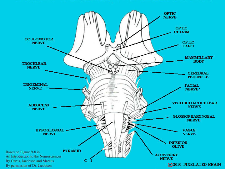
BRAINSTEM and CRANIAL NERVES, VENTRAL VIEW
This is the logical time to identify the cranial nerves. We don't want to get into their details here, because we take the nerves up later in the course, but (for this module) you should be able to pick out each nerve and relate its point of exit from the brainstem to surrounding structures. This figure and the next give you all you need to know, but DiganatA_5.10 is a view of them on a real brain.
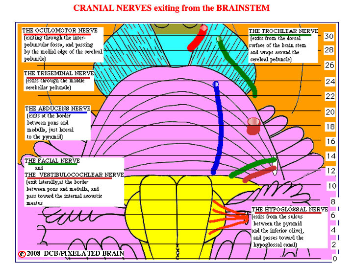
BRAINSTEM and CRANIAL NERVES, VENTRAL VIEW
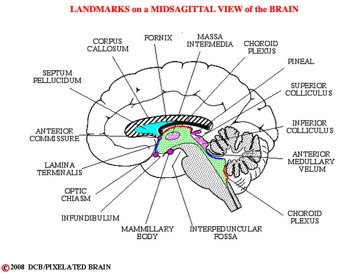
BRAINSTEM MID-SAGITTAL VIEW
Mid-sagittal (lateral) views can also be helpful. Compare with DiganatA_5.2.
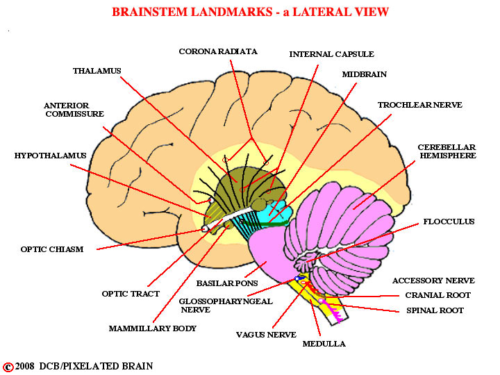
BRAINSTEM SAGITTAL VIEW
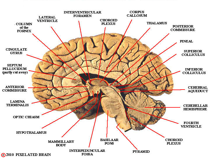
BRAINSTEM SAGITTAL VIEW
Compare with:
Diganat_5a and Diganat_5b
This completes our collection of brainstem images.
We are, in a sense, at the midpoint of the module. We have looked dorsal views of the brainstem, ventral views and sagittal ones. In the process, we have identified more than 50 structures.
To review the situation look at the Module 2 glossary. We don't expect you to know anything about the function of these structures, but we do hope you will be able to identify most of them on the appropriate figure, once you finish studying this module.
The best way to get to the glossary is to click on "I Want To" , which you will find somewhere in most views (including this one) . In that view, find the "X" for the module 2 glossary and click on it.