MODULE 2 - SECTION 6 - BRAIN MODEL

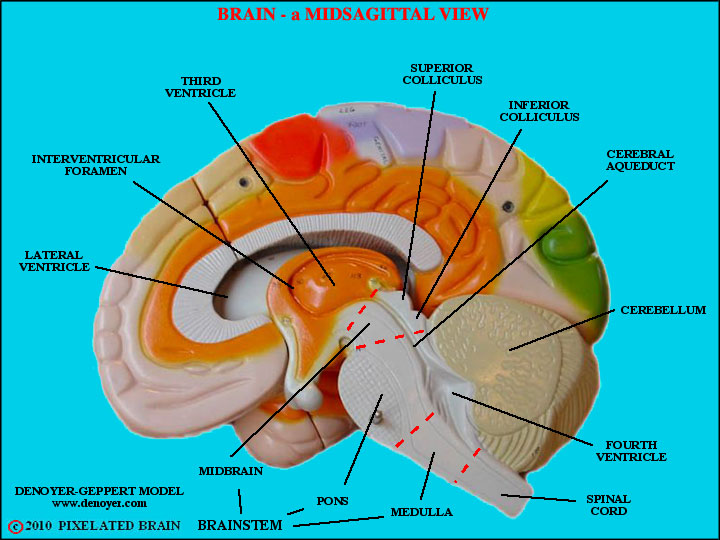
     When we looked at the big brain model, at the end of Module 1, we avoided labeling brainstem structures, partly because they don't show up very clearly on the model, but mainly because that would have meant getting ahead of ourselves. Now, we look at this model for the last time.  

     Lines mark the extent of the medulla, pons and midbrain. While you have this view on your screen you might want to call up Figure 1-40 for comparison.
1
      Shows the optic tract wrapping around the cerebral peduncle to terminate in the lateral geniculate nucleus, the trigeminal nerve exiting by passing through the middle cerebellar peduncle, the inferior olive lying just lateral to the pyramid, and more. Compare it with Figure 2-16.
2
     A dorsal view. Reminds you that the cerebellum normally obscures our view of the dorsal surface of the brainstem. Compare with Figure 2-8. Shows nicely how the trochlear nerve exits from the dorsal surface of the brainstem, just caudal (behind) the inferior colliculus and begins its long journey by passing laterally and wrapping around the cerebral peduncle. Compare, also, with Figure 2-16.
3

SITE MAP      MOD 2 HOME      PIXBRAIN HOME       I WANT TO      COMMENT

NEXT