MODULE 2 - SECTION 8 - THE SPINAL CORD
This view tries to make 3 points:
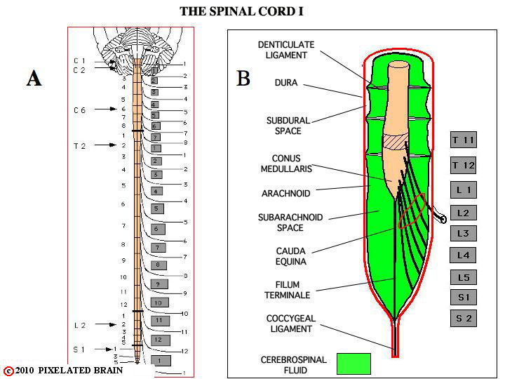
1) It shows the relative size of the brain and spinal cord
2) It demonstrates the relationship between spinal cord segments and vertebrae
3) It also illustrates the relationship between the meninges, the subarachnoid space and the exiting nerves in the lumbar region.
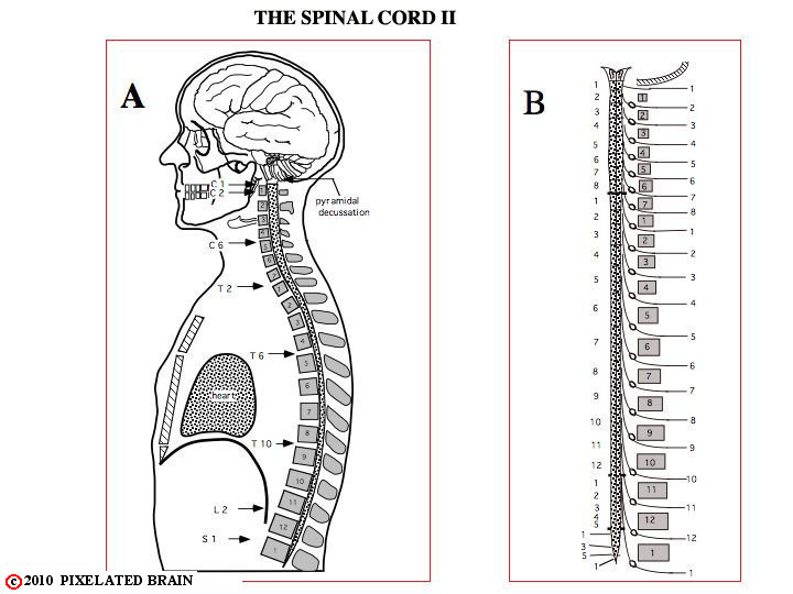
This lateral view makes some of the same points as the previous view, and relates the spinal cord to soft tissues of the thorax and the abdominal cavity.
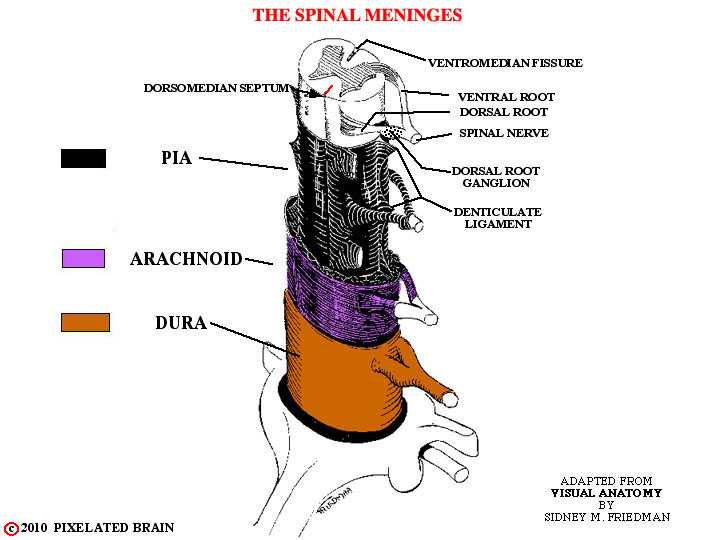
This view shows the relationship between the meninges, the subarachnoid space, the spinal cord and exiting spinal nerves.
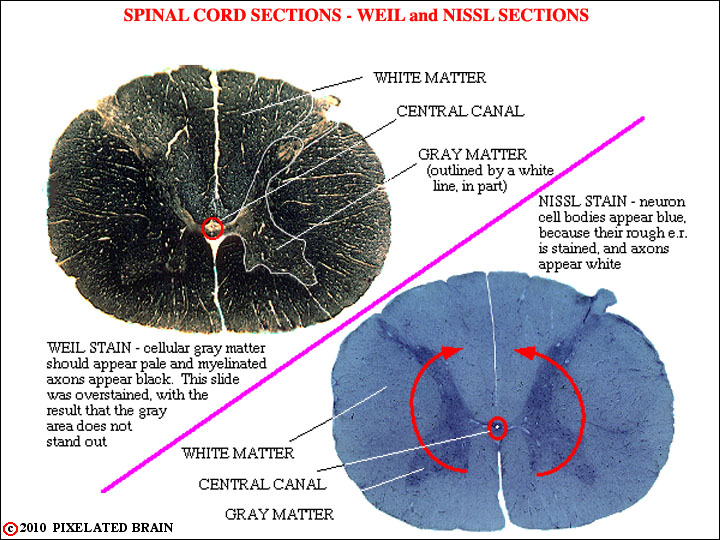
The remaining views of this section deal with the histological appearance of the spinal cord.
This first view shows the general appearance of the spinal cord.
The red arrows are there to remind you that it was the folding of the neural plate that gave rise to the neural tube, of which the spinal cord is a part.
The cavity within the tube is quite small at spinal cord levels (the central canal), but greatly expands within some parts of the brain, as you already have discovered.
At any level, the cord may be divided into a central, butterfly-shaped region - the gray matter - which contains many cells, and a peripheral zone of white matter, consisting mainly of myelinated and unmyelinated axons.
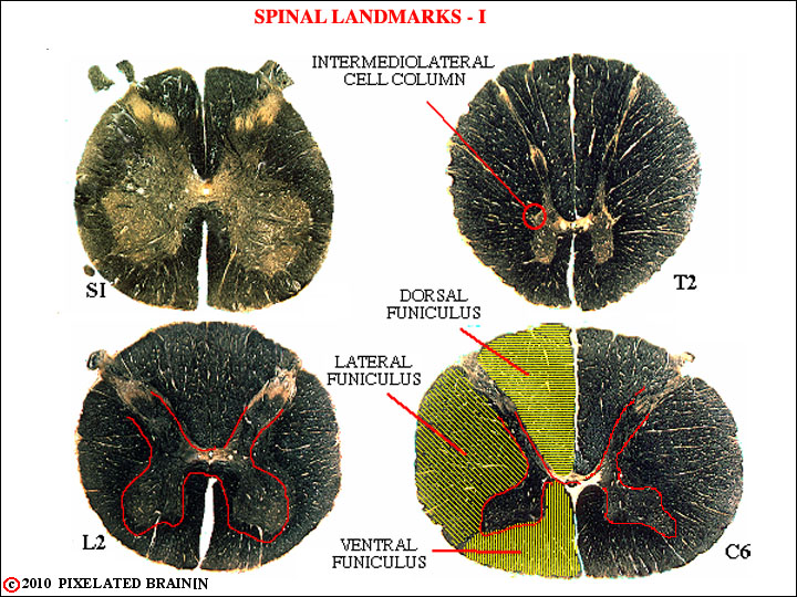
This view and the next shows several features of spinal cord anatomy that are helpful in placing the level of any given section.
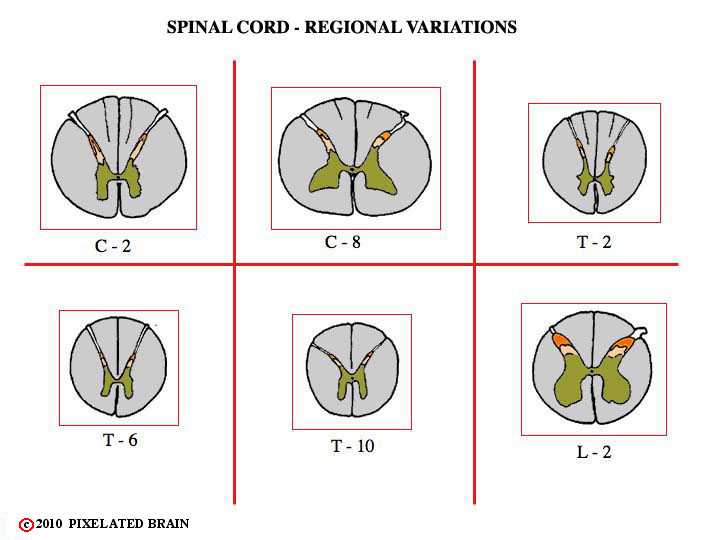
1) The gray matter enlarges at the level of the lumbosacral plexus (S1 - L2) and at the level of the brachial plexus (C5-C8)). It is relatively sparse at thoracic levels.
2) White matter occupies an increasing fraction of the total area as the cord approaches the brainstem.
3) The intermediolateral cell column is present only from T1 to L2 or L3.
- - The pathways which connect the spinal cord with the brain ascend and descend within the white matter. This peripheral region is divided by the dorsal and ventral horns of gray matter into three funiculi - the dorsal funiculus, the lateral funiculus and the ventral funiculus. At upper thoracic and all cervical levels the dorsal funiculus is further subdivided, as shown on slide 7 (2 more from here).
Another view of features of spinal cord anatomy that are helpful in placing the level of any given section.
The pathways which connect the spinal cord with the brain ascend and descend within the white matter. This peripheral region is divided by the dorsal and ventral horns of gray matter into three funiculi - the dorsal funiculus, the lateral funiculus and the ventral funiculus.
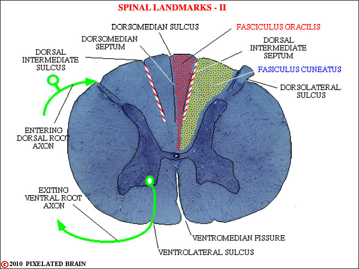
At upper thoracic and all cervical levels the dorsal funiculus is further subdivided, as shown here. This view also shows the named sulci, fissures and septi, as well as the course taken by entering and departing axons.
There are two somewhat different methods for designating regions of the spinal cord gray matter. One is based on nuclear groupings of cells (substantia gelatinosa, Clarke's nucleus, etc.) and the other divides the gray region into laminae, using a system devised by Rexed, years ago. See Blumenfeld's Figure 6.3 and Table 6.2 for a depiction of the cord and the laminae. Fitzgerald shows the laminae in his Figure 15.3 and Haines does so in his Figure 9-3. While our slides are not adequate for making out all the details on which the laminar scheme is based, they do show enough detail to make out the basic subdivisions of the cord that are of importance. You should be aware that not all authors agree on the exact assignment on names to the lamina.
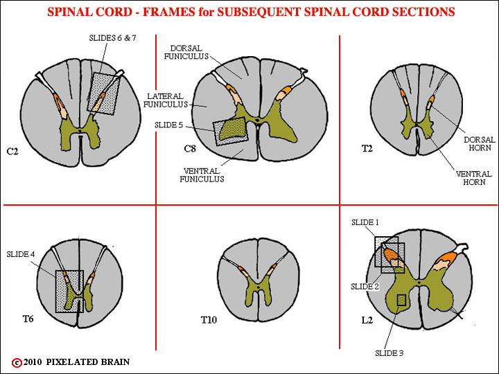
The "boxes" in this view define the areas covered in the enlarged views of the cord that follow.
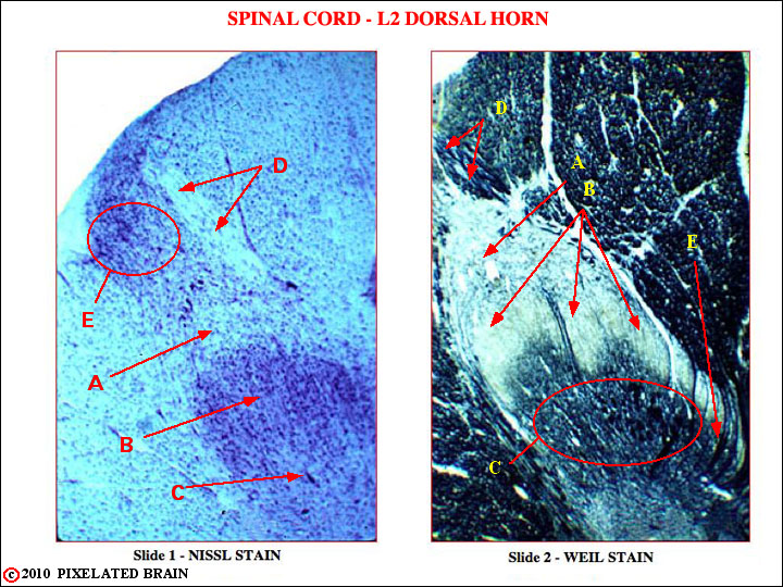
Slide 1. L2, dorsal horn. Nissl stain. Cell bodies appear pink (because their rough e. r. is stained) and axons ("white matter") appears white.
A. Posterior marginal nucleus (lamina I).
B. Substantia gelatinosa (laminae II)
C. Nucleus proprius (laminae III, IV)
D. Myelinated dorsal root fibers on their way to terminate in deeper spinal cord laminae
E. Dorsolateral fasciculus (of Lissauer). Unmyelinated dorsal root fibers enter in this more lateral position and may run up or down the cord for a few segments before turning medially to terminate in laminae I, II, III.
Slide 2. L2 dorsal horn. Weil stain. Cellular gray matter appears pale, while myelinated axons stain black. Almost same level as previous slide.
A. Posterior marginal nucleus (lamina I).
B. Substantia gelatinosa (laminae II)
C. Nucleus proprius (laminae III, IV)
D. Myelinated dorsal root fibers on their way to terminate in deeper spinal cord laminae
E. Note myelinated dorsal root axons turning laterally to terminate directly in the deeper laminae
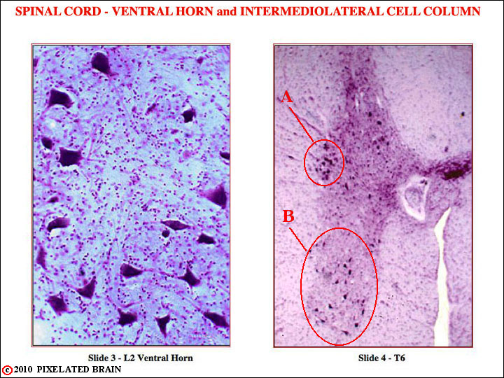
Slide 3. L2 ventral horn, higher magnification. Nissl stain. Note the large cell bodies of motor neurons in what must be lamina IX
Slide 4. T6. Nissl stain
A. Intermediolateral cell column
B. Motor neuron nuclei in ventral horn. Note that at this thoracic level, where the motor innervation is of trunk muscles only (no extremity), the anterior horn is rather narrow. Compare with the anterior horn at the level of C6, shown in Figure 2-48.
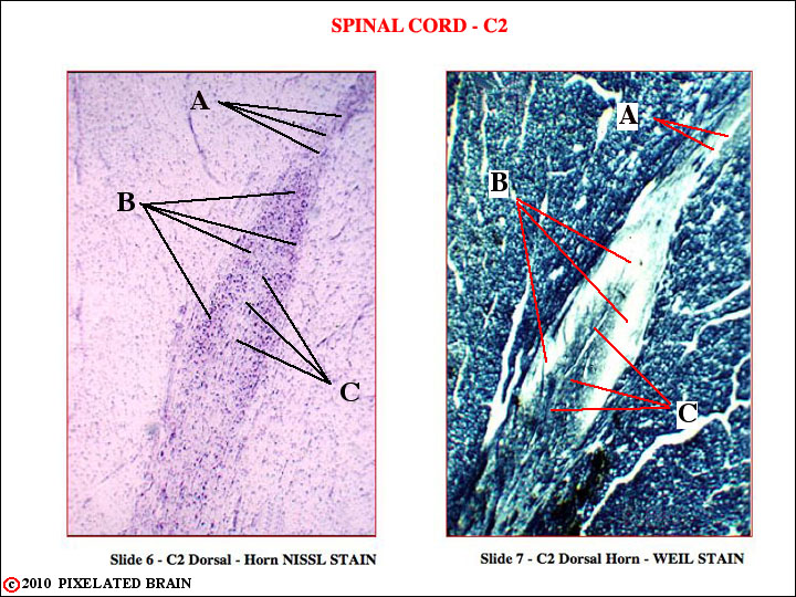
Slide 6. C2 dorsal horn, Nissl stain.
A. Posterior marginal nucleus (lamina I).
B. Substantia gelatinosa (laminae II
C. Nucleus proprius.
Slide 7. C2 dorsal horn, Weil stain.
A. Posterior marginal nucleus (lamina I).
B. Substantia gelatinosa (laminae II)
C. Nucleus proprius.
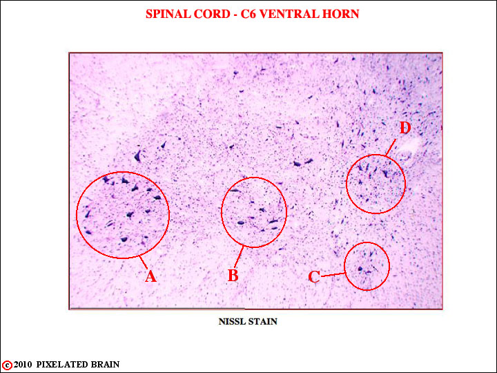
Slide 5. C6 ventral horn. Nissl stain
Note the prominent lateral extension of the ventral horn. Letters A-D point to the individual motor nuclei of lamina IX, each of which provides innervation to a single muscle or a group of muscles having a similar action. Distal muscles (i.e., those of the hand) tend to be innervated by the more lateral nuclei, such as A. Proximal muscles (like the deltoid) are innervated by the most medial nuclei, such as C and D. For diagrams illustrating this arrangement see: Blumenfeld Fig. 6.7, Fitzgerald Fig. 16.1, Haines Fig.9-8.
This type of organization, in which the body is represented spatially within the nervous system in a reliable and orderly fashion is called a somatotopic one. Somatotopy is conceptually similar to the organization in other sensory systems (retinotopy for the visual system and tonotopy for the auditory system.