MODULE 3 - SECTION 1 - AN OVERVIEW of the DC-ML
1
2
3
4
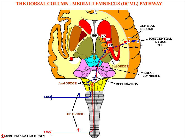
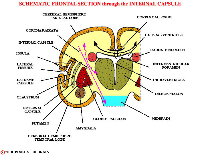
Note that on the left, the letters L and A show the position of the "arm" and "leg" fibers in the posterior limb of the capsule
5
|
MODULE 3 - SECTION 1 - AN OVERVIEW of the DC-ML |
|
|
T |
1 |
|
|
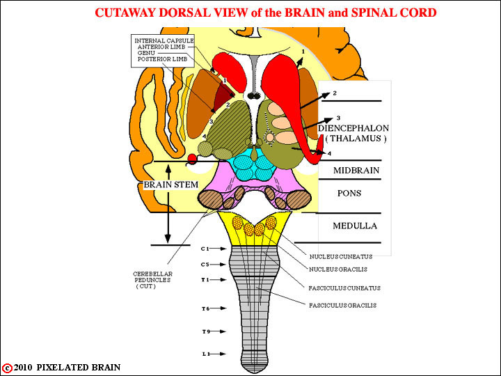
This view is a modified version of the one shown above. On the right, four "tan colored" thalamic nuclei have been added and numbered arrows show how fibers radiate laterally from the thalamus, passing "under" the caudate, and entering the internal capsule. On the left, a horizontal cut has been made through the brain, revealing the classic "V" shape of the internal capsule. The "1" on both sides shows the position of fibers traveling in the anterior limb of the capsule; The "2, 3, 4" show the position of fibers traveling in the posterior limb. |
2 |
|
|
T |
3 |
|
|
T |
4 |
|
|
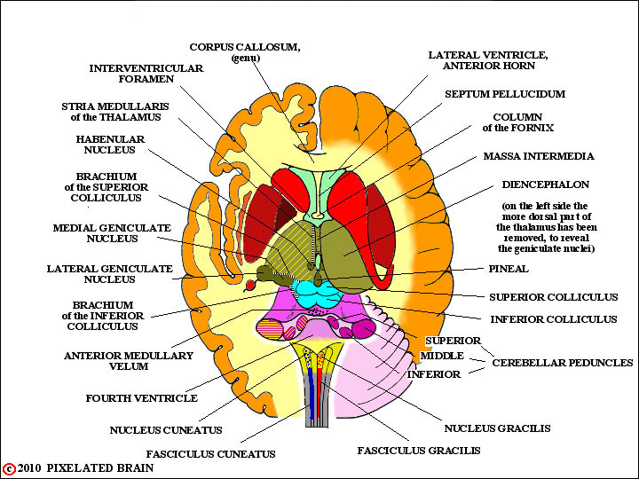
This view simply draws in the DC-ML pathway on our standard dorsal view of the brainstem and thalamus. Note that the pathway retains a somatotopic plan of organization all the way up to the cortex. VP is an abbreviation for ventralis posterior, the name of the thalamic nucleus where the cell bodies of the third order neurons are found.
Note that on the left, the letters L and A show the position of the "arm" and "leg" fibers in the posterior limb of the capsule |
5 |
|