MODULE 3 - VIEWS-ALL 1
The
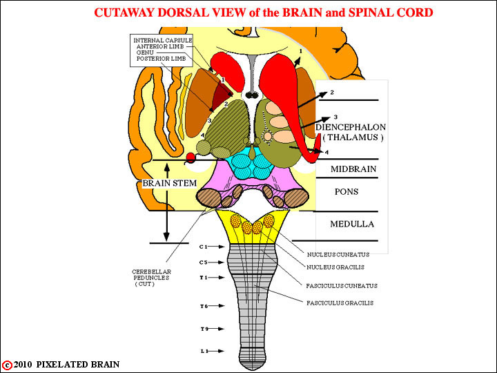
FIGURE 3-1
This view is similar to that of Figure 2-4, except that the numbers denoting slide levels have been removed and labels showing the regions of the brainstem (and the diencephalon) have been added. In addition, numbers and arrows have been added to help us describe the internal capsule. On the right, the numbered arrows show how fibers radiate laterally from the thalamus, passing "under" the caudate, and entering the internal capsule. On the left, a horizontal cut has been made through the thalamus, revealing the classic "V" shape of the internal capsule. The "1" on both sides shows the position of fibers traveling in the anterior limb of the capsule; The "2, 3, 4" show the position of fibers traveling in the posterior limb.
Note the position of the nuclei and fasciculi associated with the DC-ML pathway.
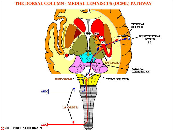
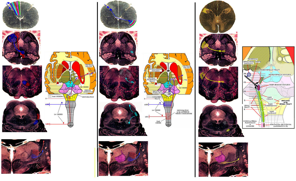
FIGURE 3-2
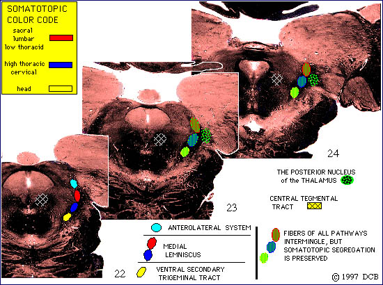
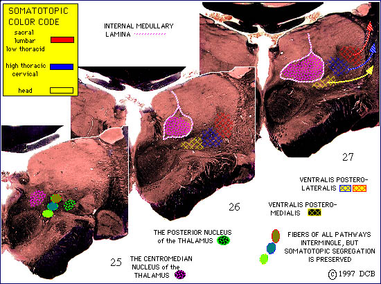
This view simply draws in the DC-ML pathway on our standard dorsal view of the brainstem and thalamus. Note that the pathway retains a somatotopic plan of organization all the way up to the cortex. VP is an abbreviation for ventralis posterior, the name of the thalamic nucleus where the cell bodies of the third order neurons are found.
Note that on the left, the letters L and A show the position of the "arm" and "leg" fibers in the posterior limb of the capsule.

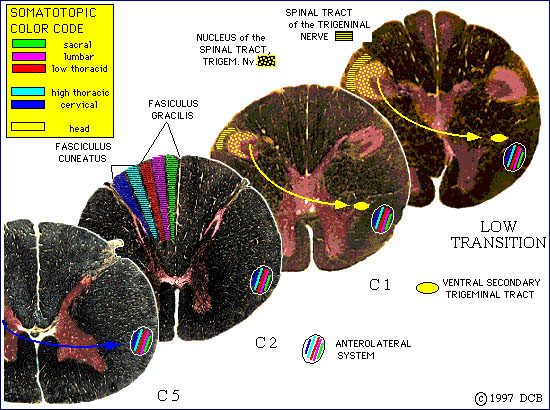
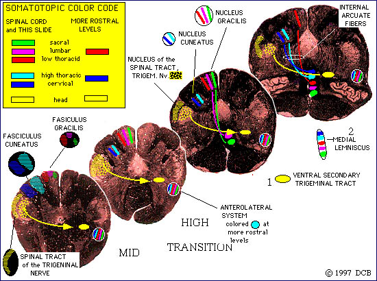
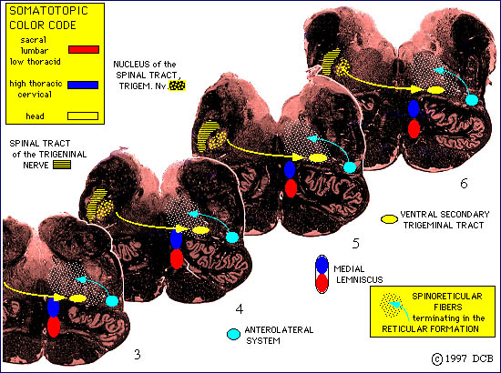
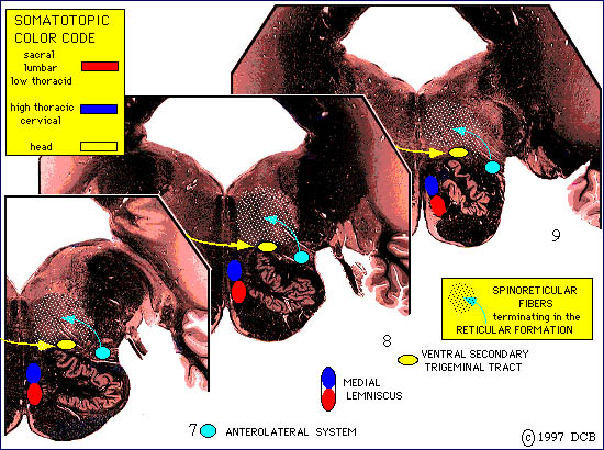
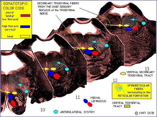
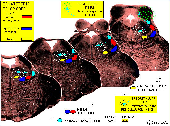
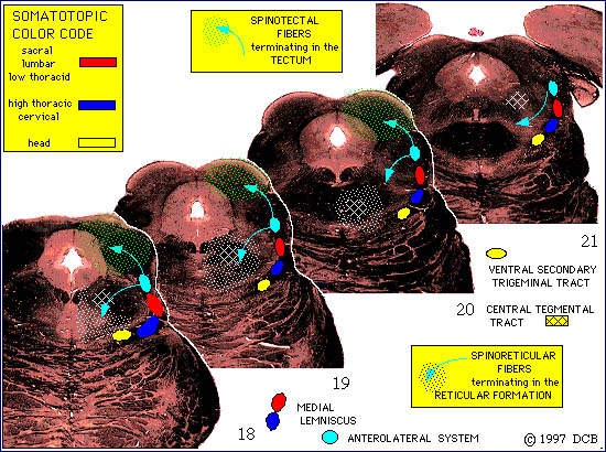
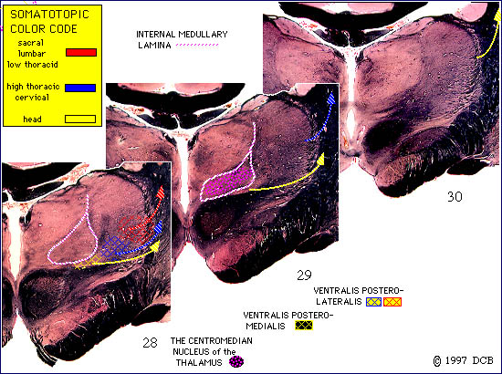
Fibers of this pathway are organized in a somatotopic manner (fibers from adjacent parts of the body's surface run in adjacent parts of the pathway), and the color code used to represent this is:
Sacral - green
Lumbar - purple
Low Thoracic - red
High Thoracic - light blue
Cervical - dark blue
The first order neurons of this pathway are large myelinated axons which enter in the medial part of the dorsal root. The region where each dorsal root enters the spinal cord is often called the "dorsal root entry zone" and these fibers enter in the medial part of the zone. The fibers then bifurcate, with one branch passing deeply to terminate in the dorsal horn and the other branch passing medially to ascend in the posterior funiculus. Because of the way in which the ascending fibers pile up in the posterior funiculus, we say that it is somatotopically organized, with the lower part of the body represented most medially.
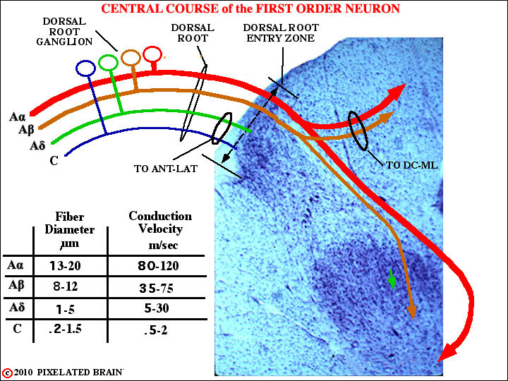
FIGURE 3-4
This view simply shows how a typical axon of the DC-ML pathway enters the cord. One branch (D) passes deeply to terminate in the dorsal horn. A second branch passes medially to enter the posterior funiculus (= dorsal column). For identification of the letters see Figure 2-34.
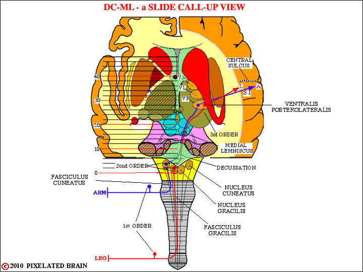
FIGURE 3-5
Sections
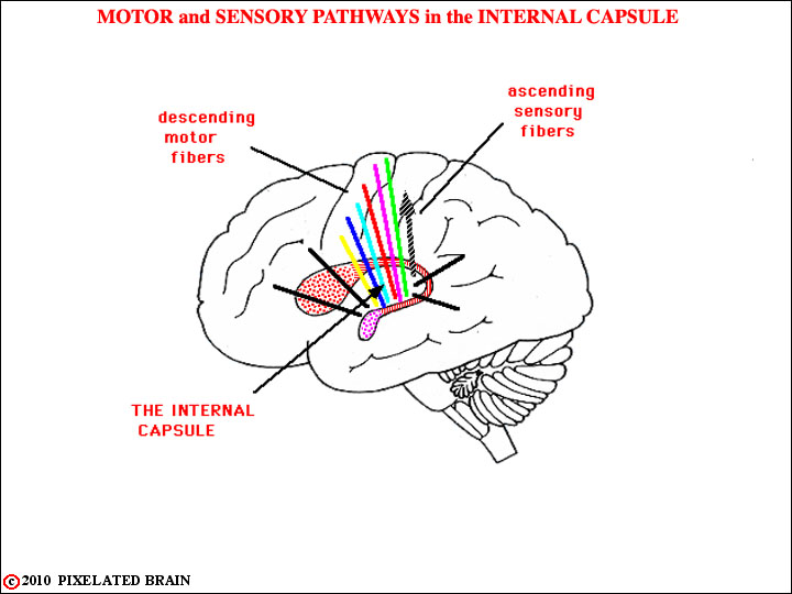
FIGURE 3-6
This is a lateral view of the brain (we are looking medially), with the putamen and globus pallidus (together = the lentiform nucleus) removed. Take a look at Figure 1-42 to remind yourself of the relationships between all these structures. Because of what has been removed we can see the fibers of the internal capsule. The colored ones are part of the descending motor system, but just posterior to them a single shaded arrow shows the course taken by the ascending fibers of the somatic sensory system that we have been tracing.
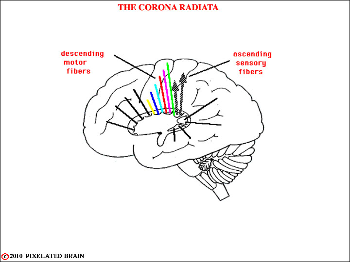
FIGURE 3-7
This view is similar to the previous one, but now the lentiform nucleus has been put back in place so we don't see the ascending somatic sensory fibers until they emerge above the dorsal edge of the nucleus. Here, they are said to form a part of the corona radiata. Note hat they are headed for the post central gyrus.
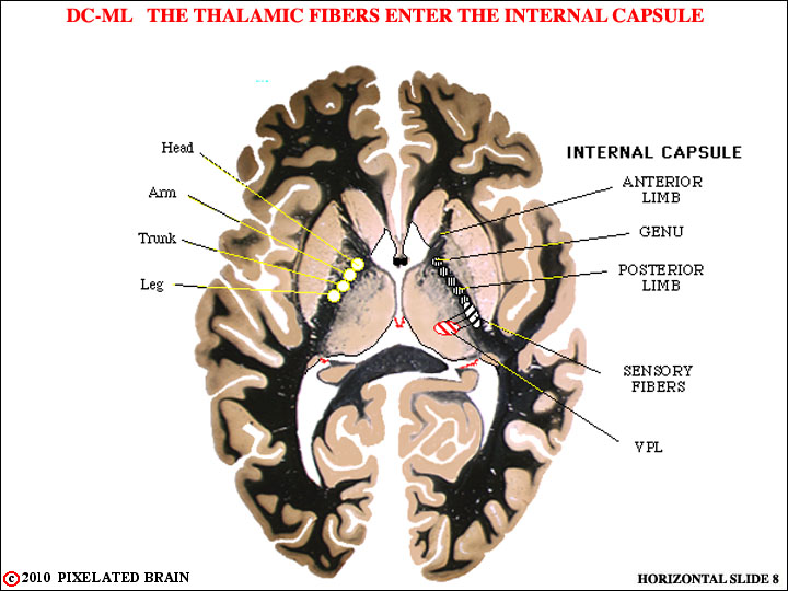
FIGURE 3-8
This is a horizontal section that shows the "classic" V shape of the internal capsule. The position of the nucleus ventralis posterolateralis (VPL) has been drawn in and you can see the course of fibers passing laterally from it to enter the more posterior part of the posterior limb of the internal capsule.
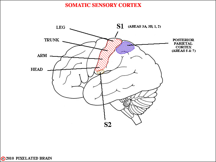
FIGURE 3-9
The ascending fibers of the DC-ML system terminate in cortical area S1. Like the pathway, this cortical receiving area is somatotopically organized. From S1 there are relays to S2 and to the posterior parietal cortex. See your lecture notes for greater detail.
FIGURE 3-10
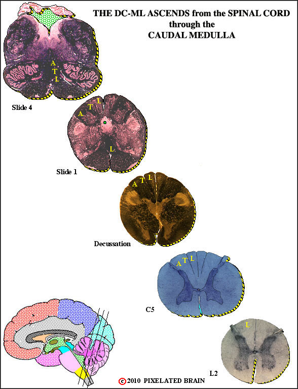
FIGURE 3-11
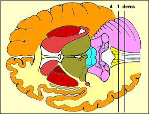
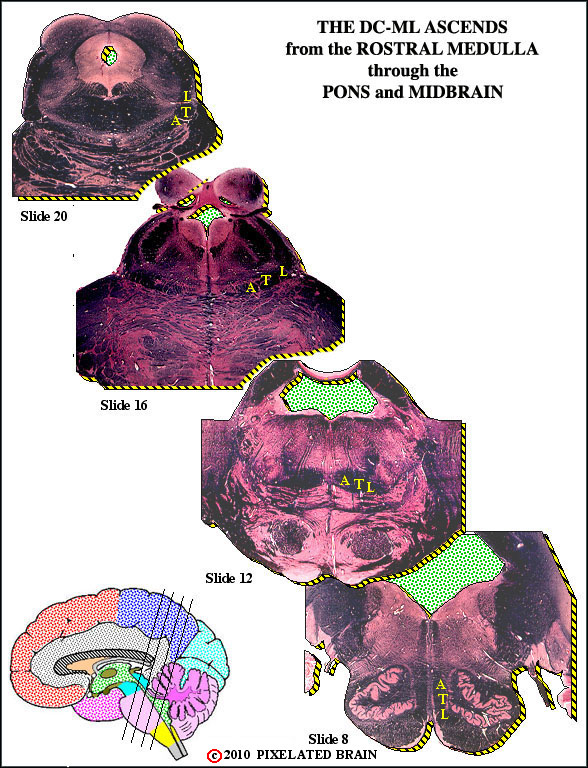
FIGURE 3-12
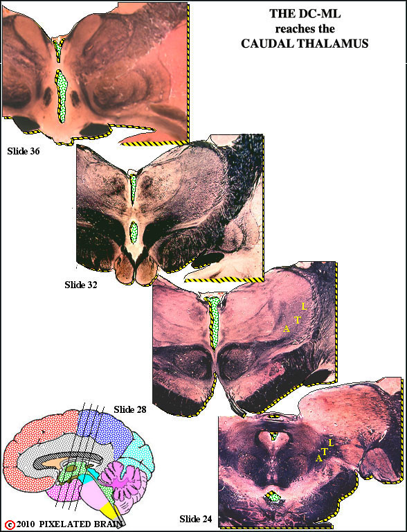
FIGURE 3-13
FIGURE 3-14
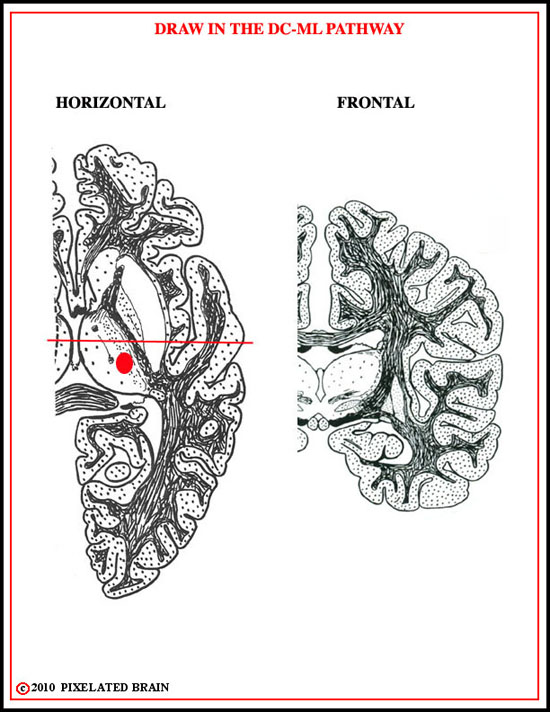
FIGURE 3-15
FIGURE 3-16
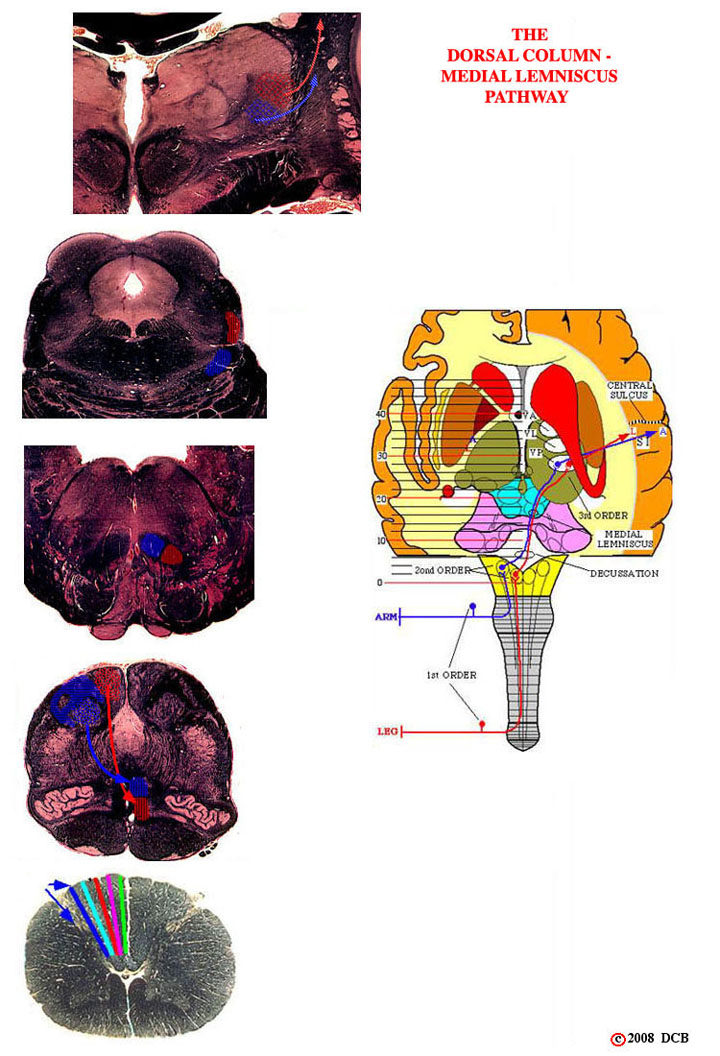
FIGURE 3-17