MODULE 5 - VIEWS-ALL
The

FIGURE 5-1
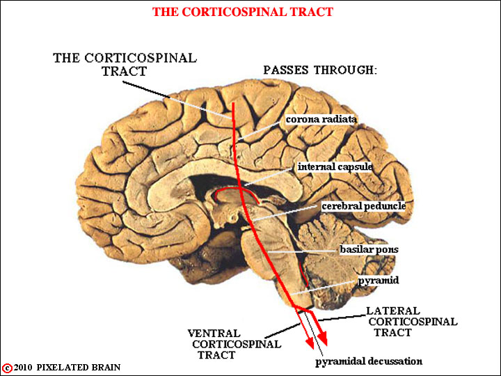
FIGURE 5-2
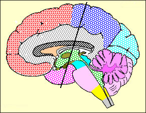
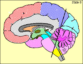
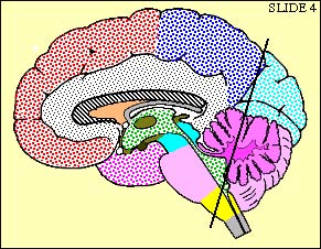
Shown in red is the course of the descending corticospinal (pyramidal) tract, projected on a midsagittal view of the gross brain.
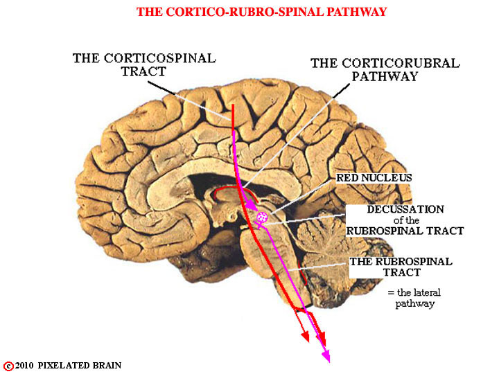
FIGURE 5-3
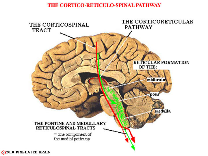
FIGURE 5-4
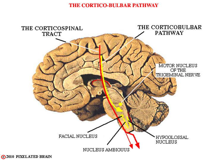
FIGURE 5-5
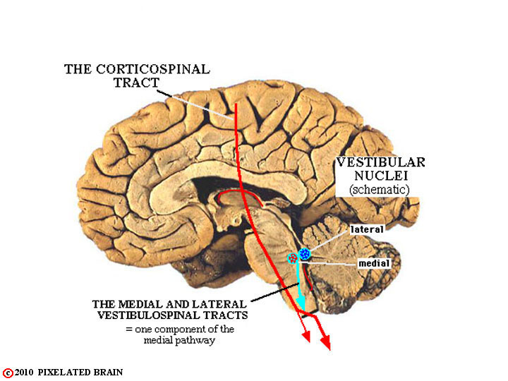
FIGURE 5-6
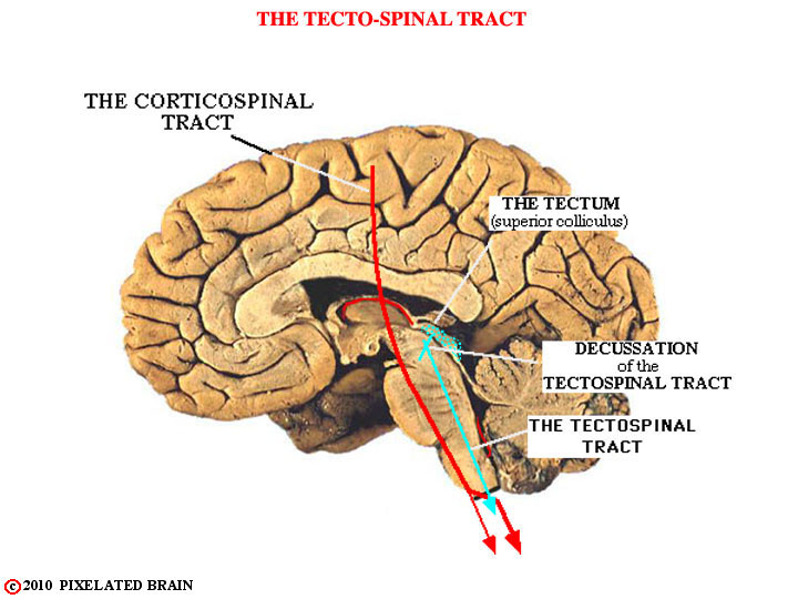
FIGURE 5-7
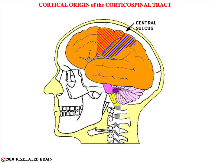
FIGURE 5-8
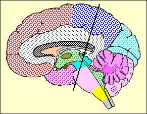
The corticospinal tract (all those descending fibers that pass through the pyramids of the medulla) originate in a broad area lying in front of and behind the central sulcus. That part of the tract which starts out in the parietal lobe (blue) appears to be concerned with modulating sensory input rather than initiating movement and it will not be considered in detail in this module. The precentral component of the tract - the part responsible for motor function - is organized in a somatotopic manner at most levels.
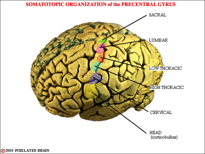
FIGURE 5-9
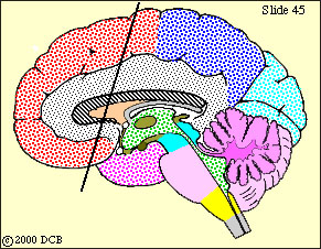
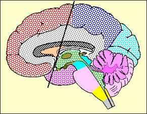
This view shows the color scheme that will be used in subsequent views.
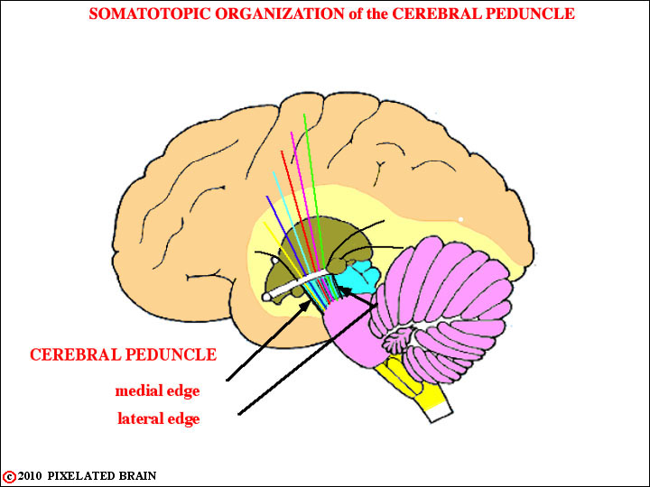
FIGURE 5-10
This view, and some that follow, tries to show how the somatotopic order at the cortical level is reversed at the level of the cerebral peduncle. At the cortical level, "head" fibers originate laterally and "leg" fibers originate medially. In the peduncle "head" fibers are in the most medial part of the tract and "leg" fibers in the most lateral part of it.
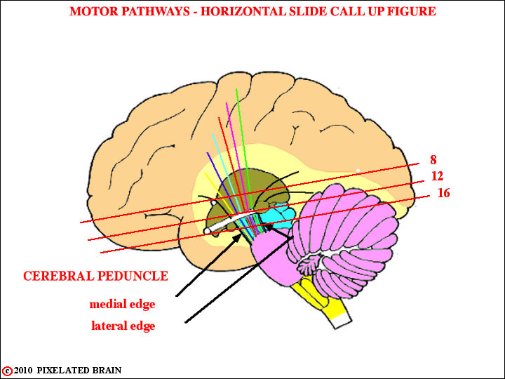
FIGURE 5-11
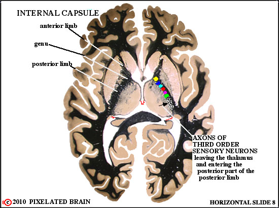
SOMATOTOPIC ORGANIZATION of_MOTOR SYSTEM in INTERNAL CAPSULE
Corticobulbar or "head" fibers pass through the genu of the internal capsule, and the fibers of the corticospinal tract pass through the anterior part of the posterior limb. Compare with MRI AXIAL view of Blumenfeld Figure 4.13G.
SOMATOTOPIC ORGANIZATION of the MOTOR SYSTEM
 All these descending fibers have passed around the thalamus, and are headed caudally, to reach the ventral surface of the midbrain. Compare with Blumenfeld Figure 4.13F (the level is slightly different).

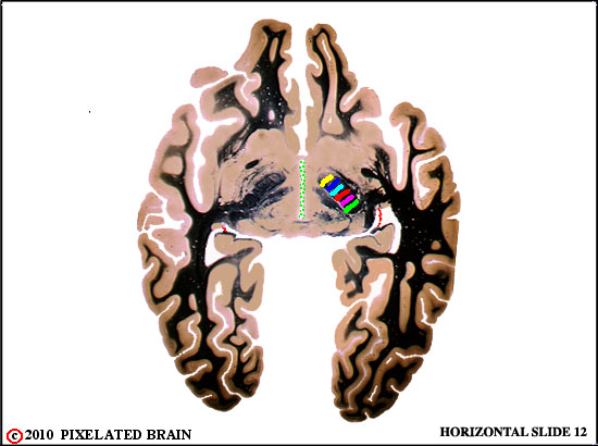
SOMATOTOPIC ORGANIZATION of_MOTOR SYSTEM in CEREBRAL PEDUNCLE Both the corticospinal tract and the corticobulbar pathway now lie within the cerebral peduncle. Compare with Blumenfeld Figure 4.13E.
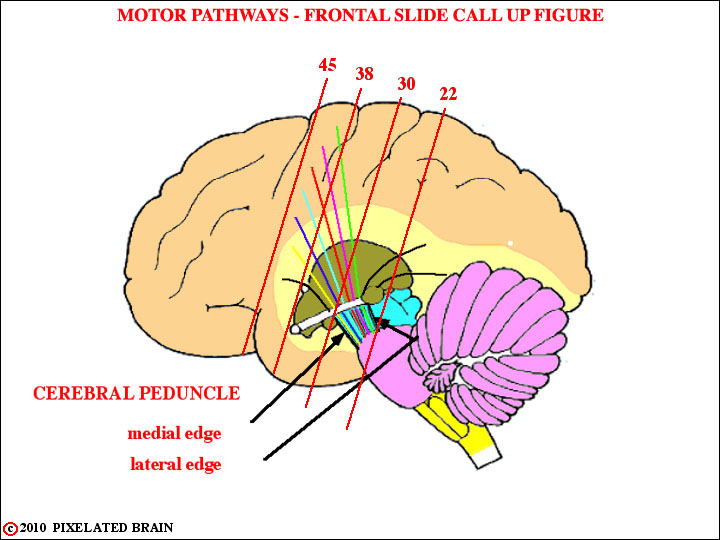
FIGURE 5-12
CORTICAL ORIGIN of the CORTICOSPINAL TRACT
Here we see fibers leaving the cortex and descending through the white matter of the hemisphere.
THE CORTICOSPINAL TRACT enters the INTERNAL CAPSULE
Descending fibers pass through the corona radiata, into the internal capsule.
THE CORTICOSPINAL TRACT in the INTERNAL CAPSULE
Fibers move ventrally and caudally, around the thalamus, to approach the cerebral peduncle on the ventral surface of the midbrain.

THE CORTICOSPINAL TRACT in the CEREBRAL PEDUNCLE
Fibers descend through the cerebral peduncle and begin to enter the basilar pons.
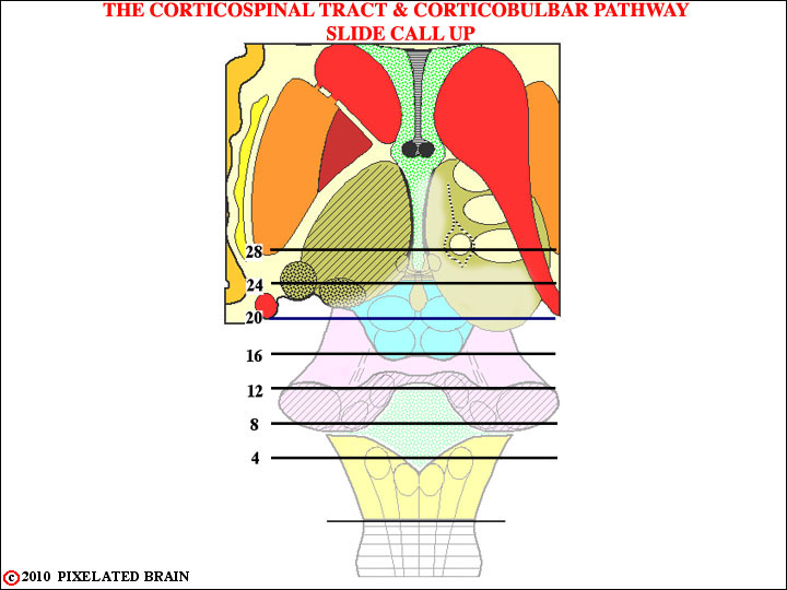
FIGURE 5-13
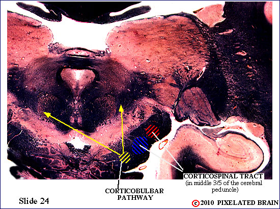
CORTICOSPINAL TRACT enters the CEREBRAL PEDUNCLE
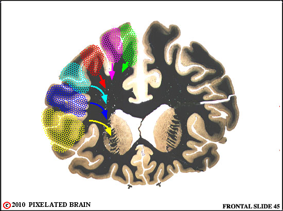
Fibers have descended around the thalamus in the internal capsule and are now beginning to emerge on the ventral surface of the brain as they enter the cerebral peduncle. Note the somatotopic organization. Corticobulbar fibers are beginning to depart and move dorsally, descending diffusely through the tegmentum of the brainstem. Note the oculomotor nerve, exiting from the interpeduncular fossa, just medial to the cerebral peduncle. Some of the thalamic nuclei, in the more dorsal part of this slide, should look familiar to you.
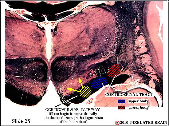
CORTICOSPINAL TRACT in the CEREBRAL PEDUNCLE
Corticobulbar fibers continue to leave; those long yellow arrows are meant to show the general course the fibers take. Some will stay on the ipsilateral side of the brainstem and others will cross the midline to descend on the contralateral side. The corticospinal tract occupies the middle part of the cerebral peduncle. While the plane of the slide is oblique, the ventral part of it passes through the midbrain.
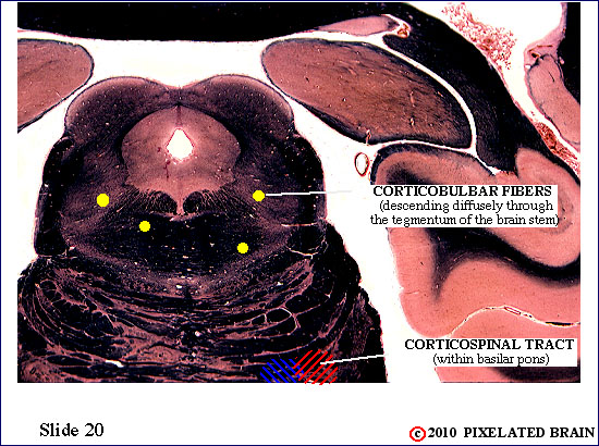
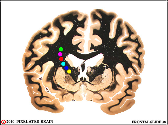
CORTICOSPINAL TRACT in the BASILAR PONS
The little yellow dots are our attempt to remind you that corticobulbar fibers are descending diffusely through the tegmentum of the brainstem. The corticospinal fibers are now buried in the basilar pons.
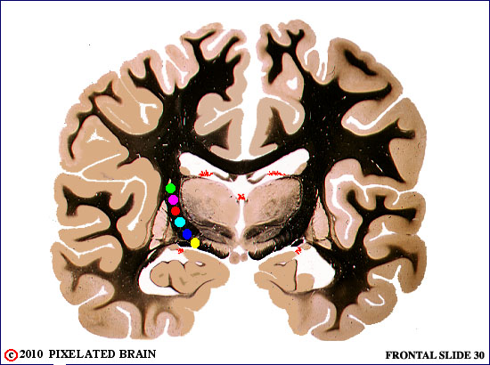
20
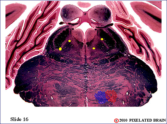
CORTICOSPINAL TRACT in the BASILAR PONS
The pathways we are following continue their descending course. Try to identify a few landmarks as you look at each slide. For example, what are those odd, round things on the dorsal surface of the brainstem? Their presence tells us that the dorsal part of the slide passes through what division of the brainstem? There seems to be a nerve exiting from the brain just below them; which one is it?
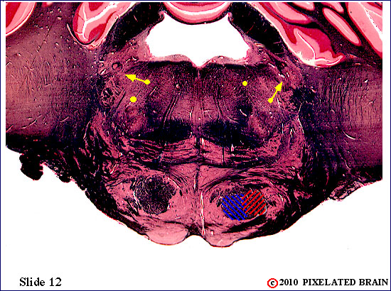
CORTICOSPINAL TRACT in the BASILAR PONS
The corticospinal fibers are still in the basilar pons, and the corticbulbar ones in the tegmentum (of the pons). A look at the view of slide 12 of the Brainstem Atlas tells you that this slide passes through the motor nucleus of the trigeminal nerve. Within this nucleus are the "lower motor neurons" that innervate the muscles of mastication - the ones you use to chew and bite. The neurons of this nucleus must receive an input from "upper motor neurons" within the "head" area of the motor cortex. We have been representing the descending axons of these neurons by the yellow dots, and now some of them will terminate within the motor nucleus of the trigeminal nerve. Unlike the corticospinal pathway, which is almost entirely crossed, the corticobulbar one has both crossed and uncrossed components. Think, a bit, about just what this means, in terms of brain function and the effect of lesions of the corticbulbar pathway.
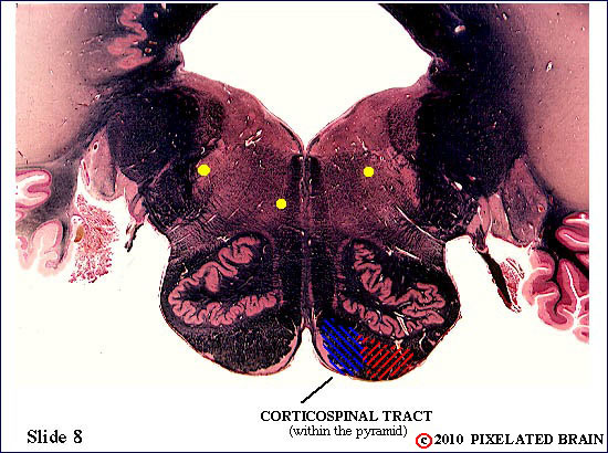
CORTICOSPINAL TRACT in the PYRAMID
The corticospinal tract has now emerged from the basilar pons and is passing through the pyramid, on the ventral surface of the medulla. Note that the corticospinal tract and the medial lemniscus now lie close together
CORTICOSPINAL TRACT in the PYRAMID
This is the last slide in which we will see those yellow dots. Why? Take a guess where some of them will terminate.

CORTICOSPINAL TRACT at the level of the PYRAMIDAL DECUSSATION
Here's where the all important decussation takes place.
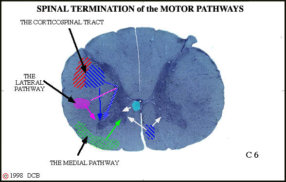
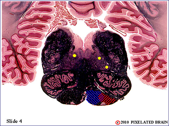
SPINAL TERMINATION of the MOTOR PATHWAYS
This slide tries to show that most corticspinal fibers terminate in the grey matter of the cord (laminae IV - IX). Via interneurons, they influence lower motor neurons in the more lateral part of the anterior horn. A few axons (the solid arrows you can barely see) terminate directly on lower motor neurons.
The rubrospinal tract is labeled the "lateral pathway" here, following the Lawrence and Kuypers model (Figure 5-1). It terminates (mostly via internuncials) in the lateral part of the anterior horn, and is mainly involved in motor control of the distal parts of the extremities. In contrast, the medial pathways terminate in the medial part of the anterior horn; they activate lower motor neurons innervating trunk and proximal extremity muscles.
FIGURE 5-14

FIGURE 5-15
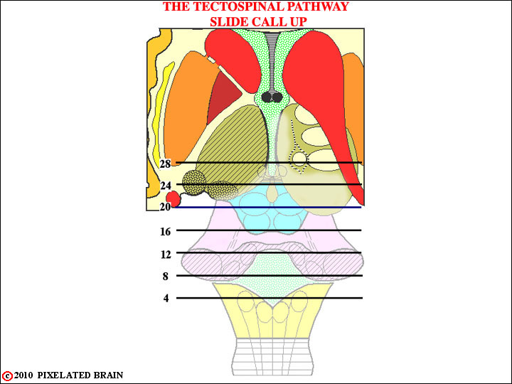
FIGURE 5-16
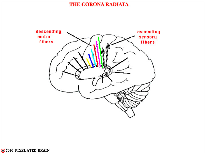
FIGURE 3-7
This view is similar to the previous one, but now the lentiform nucleus has been put back in place so we don't see the ascending somatic sensory fibers until they emerge above the dorsal edge of the nucleus. Here, they are said to form a part of the corona radiata. Note hat they are headed for the post central gyrus.
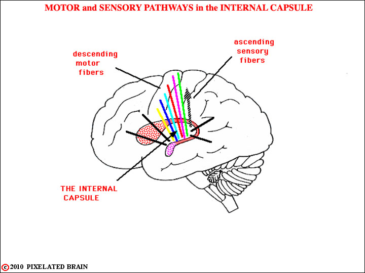
FIGURE 3-6
This is a lateral view of the brain (we are looking medially), with the putamen and globus pallidus (together = the lentiform nucleus) removed. Take a look at Figure 1-42 to remind yourself of the relationships between all these structures. Because of what has been removed we can see the fibers of the internal capsule. The colored ones are part of the descending motor system, but just posterior to them a single shaded arrow shows the course taken by the ascending fibers of the somatic sensory system that we have been tracing.