MODULE 6 - SECTION 2 - THE CORTICO-CEREBELLAR LOOP
The role of the cerebellum in planning and guiding movements is best described in terms of processes that take place sequentially.
Perhaps 100 msec. before the onset of a movement, neural messages, presumably involving the nature of the desired movement, are sent (at least, potentially) from all four lobes of the cerebral hemisphere to the cerebellum.
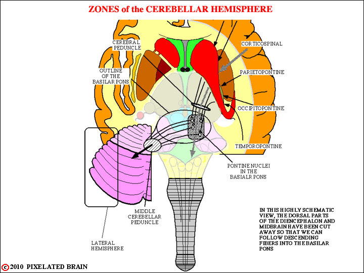
Axons of the neurons involved pass successively through the corona radiata, the internal capsule and the cerebral peduncle to reach the basilar pons. It is a massive projection and - as with most pathways of this sort - it is orderly in nature. Fibers from the frontal lobe (the fronto-pontine pathway) pass through the anterior limb of the internal capsule and then through the medial one fifth of the cerebral peduncle to reach the basilar pons. Fibers from the remaining three lobes pass through the posterior limb of the internal capsule and the lateral one fifth of the cerebral peduncle to reach the same destination. Within the cerebral peduncle these two groups of fibers are separated by the corticospinal tract, occupying the middle three fifths of the peduncle.
Within the basilar pons the descending fibers from all four lobes terminate by synapsing upon the cell bodies of the nuclei scattered diffusely throughout this region - the so-called pontine nuclei.
These neurons relay information to the cerebellum by giving rise to axons which cross the midline, run through the middle cerebellar peduncle and make synaptic contact with granule cells in the cortex of the lateral part of the cerebellar hemisphere.
Because of their appearance these axons are called mossy fibers and their specialized region of contact with granule cells is termed a glomerulus.
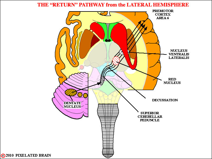
After neural processing of this input at the level of the cerebellar cortex, purkinje cells relay the output to the dentate nucleus, the largest of the "deep" nuclei, buried within the white matter of the cerebellar hemisphere.
This output is entirely inhibitory, and thus acts by modulating the tonic activity of neurons within the dentate nucleus.
Neurons within the dentate nucleus have axons which begin the return route to the the cerebral cortex. These fibers exit from the cerebellum in the superior cerebellar peduncle, wrap around the central gray region of the brain stem tegmentum, and cross the midline at the midbrain level to enter the contralateral red nucleus. A few terminate in the rostral, parvocellular division of the nucleus but the majority continue without synapsing and terminate in the nucleus ventralis lateralis of the thalamus.
The thalamic neurons, in turn, relay the message back to the cerebral cortex by way of fibers that pass through the internal capsule.