MODULE 6 - SECTION 6 - OUTPUTS PATHWAYS from the CEREBELLUM
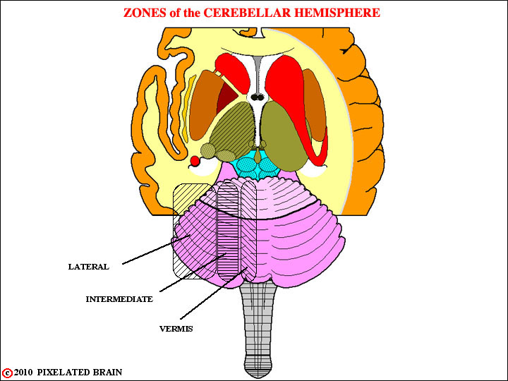
ZONES of the CEREBELLAR HEMISPHERE
The cerebellum is divided into lateral, intermediate and vermal zones. Each projects to its own set of deep nuclei, and the function carried out by each zone is distinct from that of the others. This view shows these divisions, drawn on a surface view of the cerebellum.
1. The lateral hemisphere or cerebrocerebellum, which receives its input from the contralateral cerebral hemisphere, projects back to this same structure and is involved in motor planning.
2. The intermediate (paramedian) region or spinocerebellum which receives inputs both from the contralateral cerebral hemisphere and the ipsilateral spinal cord. This region is concerned with the adjustment of motor activity involving the distal parts of the body
3. The midline region or vermis. This region actually carries out two rather different functions. It shares with the intermediate region the job of adjusting motor activity, in this case involving proximal and axial muscles. Parts of the vermis, however, have reciprocal connections with the vestibular nuclei. This subdivision of the vermis is termed the vestibulocerebellum and is concerned with monitoring and adjusting vestibular reflexes.
Each region of the cerebellar cortex projects out of the cerebellum by way of its own "deep" nucleus.
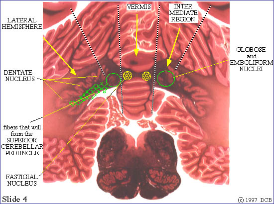
PROJECTIONS from the CEREBELLAR CORTEX to the DEEP NUCLEI
Another look at the lateral, intermediate and vermal zones of the cerebellum. Each projects to its own set of deep nuclei, and the function carried out by each zone is distinct from that of the others.
Note that the lateral part of the hemisphere (the cerebrocerebellum) projects to the dentate nucleus.
The intermediate part of the hemisphere (one component of the spinocerebellum) projects to the globose and emboliform nuclei. These nuclei, together, are also called the interposed nuclei.
After relay in these deep nuclei, fibers of both pathways depart from the cerebellum in the superior cerebellar peduncle and continue into its rostral extension, across the midline as the dentato-rubro-thalamic pathway.
As stated for previous slide:
_1. The lateral hemisphere or cerebrocerebellum, which receives its input from the contralateral cerebral hemisphere, projects back to this same structure and is involved in motorplanning.
2. The intermediate (paramedian) region or spinocerebellum which receives inputs both from the contralateral cerebral hemisphere and the ipsilateral spinal cord. This region is concerned with the adjustment of motor activity involving the distal parts of the body.
3. The midline region or vermis. This region actually carries out two rather different functions. It shares with the intermediate region the job of adjusting motor activity, in this case involving proximal and axial muscles. Parts of the vermis, however, have reciprocal connections with the vestibular nuclei. This subdivision of the vermis is termed the vestibulocerebellum and is concerned with monitoring and adjusting vestibular reflexes.
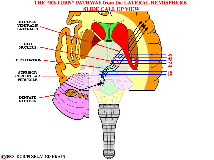
Refer to this guide to the planes of the following slides.
Use them to follow the path forward to its decussation and the red nucleus.
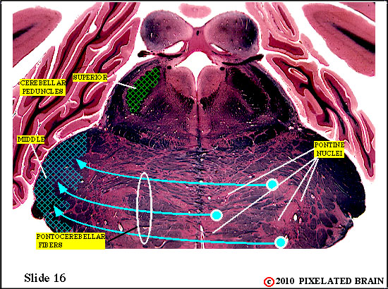
Slide 16
THE SUPERIOR CEREBELLAR PEDUNCLE ASCENDS to the MIDBRAIN
The superior cerebellar peduncle, carrying fibers from the dentate, globose and emboliform nuclei, has now left the cerebellum and is passing ventrally through the tegmentum of the brainstem.
Ignore the blue lines, showing the formation of the middle cerebellar peduncle.
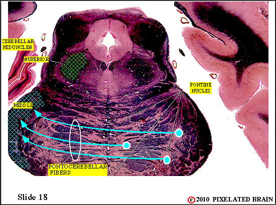
Slide 18
THE SUPERIOR CEREBELLAR PEDUNCLE ASCENDS to the MIDBRAIN
The superior cerebellar peduncle, carrying fibers from the dentate, globose and emboliform nuclei, is passing ventrally through the tegmentum of the brainstem.
Ignore the blue lines, showing the formation of the middle cerebellar peduncle.
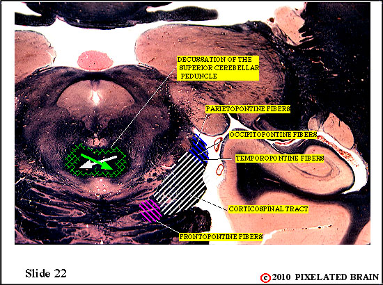
Slide 22
DECUSSATION of the SUPERIOR CEREBELLAR PEDUNCLE
The superior cerebellar peduncle, carrying fibers from the dentate, globose and emboliform nuclei, now decussates
Ignore the fibers of the descending pathways, shown within the cerebral peduncle.
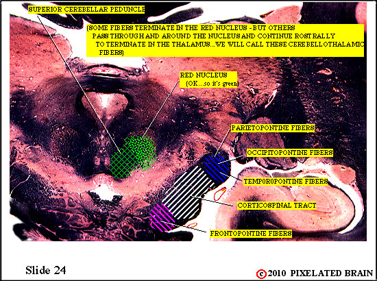
Slide 24
THE SUPERIOR CEREBELLAR PEDUNCLE enters the RED NUCLEUS
The superior cerebellar peduncle, carrying fibers from the dentate, globose and emboliform nuclei, now approaches the red nucleus.
Ignore the fibers of the descending pathways, shown within the cerebral peduncle.
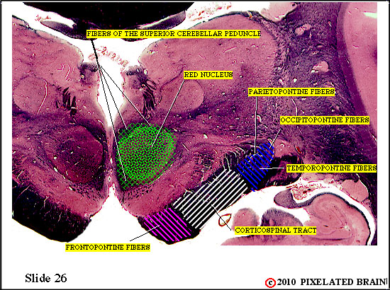
Slide 26
THE SUPERIOR CEREBELLAR PEDUNCLE enters the RED NUCLEUS
At the level of the red nucleus, the output limbs of the pathways part company. Fibers of the cortical path(the one originating in the dentate nucleus) may relay in the parvocellular part of the red nucleus but for the most part continue on to terminate in the nucleus ventralis lateralis of the thalamus (see Slides 28 & 30). From there they project to the premotor area of the cortex (Area 6). Fibers of the spinal cord path (the ones originating in the globose and emboliform nuclei) terminate in the magnocellular part of the red nucleus ending upon cells that contribute axons to the rubrospinal pathway. Thus, the rubrospinal path completes the spinal cord loop and is the route by which the intermediate part of the cerebellum can modify ongoing motor activity.
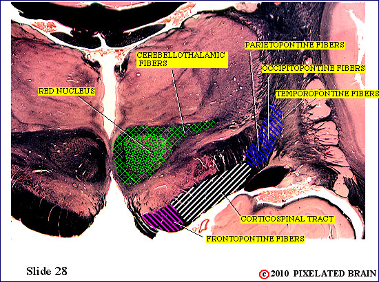
Slide 28
CEREBELLOTHALAMIC FIBERS approach the THALAMUS
Fibers of the cortical pathway continue rostrally. Here they are labeled cerebellothalamic fibers. They are moving laterally, just ventral to VPM and VPL, to approach the nucleus ventralis lateralis.
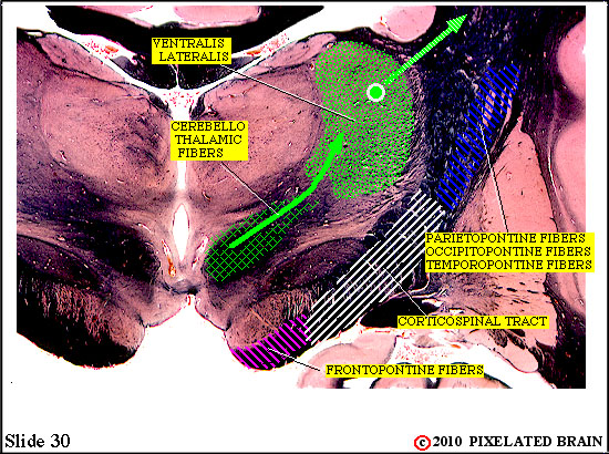
Slide 30
CEREBELLOTHALAMIC FIBERS enter VENTRALIS LATERALIS
Fibers of the cortical pathway now enter the nucleus ventralis lateralis. After a synaptic relay here, the pathway continues onward to the cortex, passing through the internal capsule.