MODULE 8 - INTRODUCTION
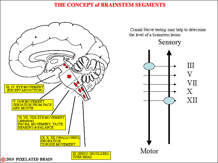
The cranial nerves carry out sensory and motor functions associated with structures in the head in much the same manner as the spinal nerves do for the body. In addition, they are concerned with the "special senses" and innervate muscles that we term "special" because of their unique embryological origin from branchial arches. Finally, lesions within the cranial cavity often manifest themselves first as a defect in some aspect of cranial nerve function. Frequently, the combination of damage to long tracts (i.e., the medial lemniscus, the pyramidal tract) and to certain cranial nerves creates a group of findings which allows the physician to fix the site of the disease with considerable precision . But to do so, it is essential to have a good three dimensional picture of where each cranial nerve is and how the information carried over the nerve originates or distributes within the brainstem. The goal of this module and the next one is to help you acquire this picture.
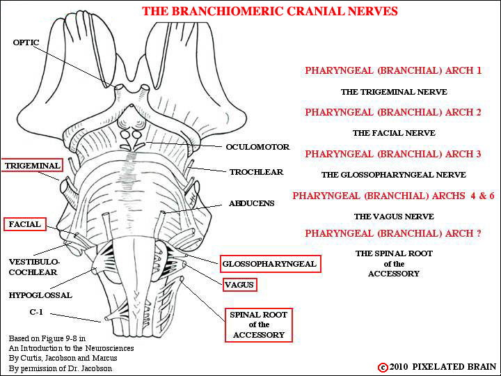
This module deals with the trigeminal nerve, the facial nerve, the glossopharyngeal nerve, the vagus nerve and the accessory nerve. These nerves have in common the fact that they provide motor innervation to muscles of branchial arch origin, and that is why we call them the "branchiomeric group" (Figure 8-2).

