MODULE 8 - SECTION 3 - AN OVERVIEW of each CRANIAL NERVE
Let's start by considering each of the five cranial nerves that are taken up in this module individually.
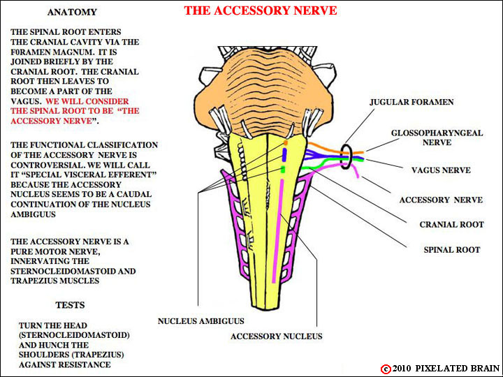
Figure 8-13
This figure gives the details of the accessory nerve. For us the accessory nerve is the structure that you can actually dissect in the neck. As this figure makes clear, it is the equivalent of the spinal accessory nerve.
You have seen the accessory nerve before, in views 2-16, 2-41, 2-42 and 2-43. Another nice view of the real thing is gluhb_01_3.html , but it is not labeled in that view.
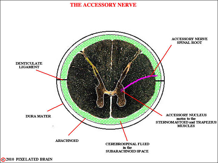
This figure shows how the nerve originates in the upper cervical cord, exits from the lateral aspect of the cord as a series of rootlets and ascends in the subarachnoid space to enter the cranial cavity by way of the foramen magnum.
THE VAGUS NERVE
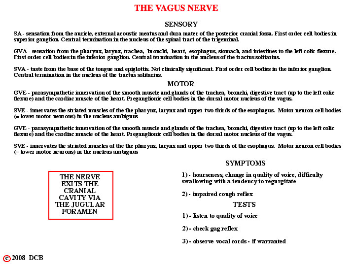
The details of the vagus nerve.
Note that We should have included chemo and baroreceptors from the aortic arch in the GVA list. All these GVA fibers terminate in the caudal part of the Nucleus of the Tractus Solitarius.
For views of the exiting Vagus Nerve, see Figure 2-16, Figure 2-39, Figure 2-41 and Figure 2-42.
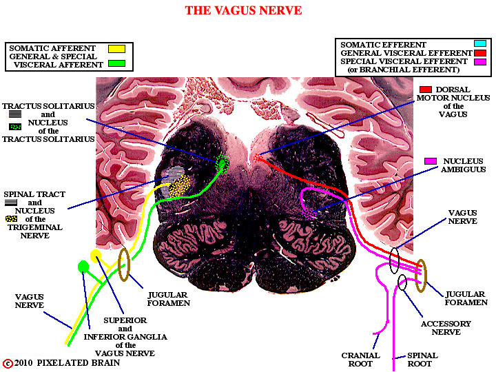
THE VAGUS NERVE
This figure hows the nuclei that give rise to the motor components of the nerve and the sensory nuclei that receive the incoming sensory fibers.
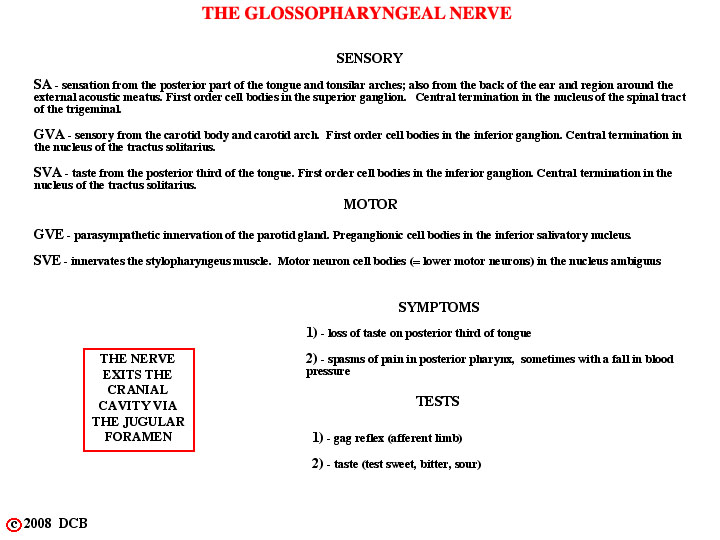
THE GLOSSOPHARYNGEAL NERVE
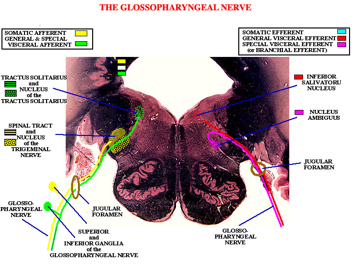
THE GLOSSOPHARYNGEAL NERVE
This shows the nuclei that give rise to the motor components of the nerve and the sensory nuclei that receive the incoming sensory fibers.
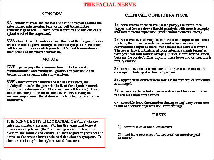
THE FACIAL NERVE
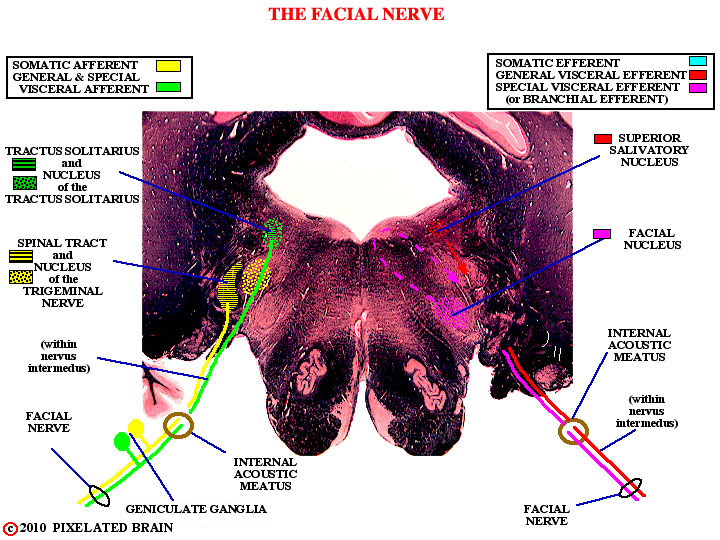
THE FACIAL NERVE
This shows the nuclei that give rise to the motor components of the nerve and the sensory nuclei that receive the incoming sensory fibers.
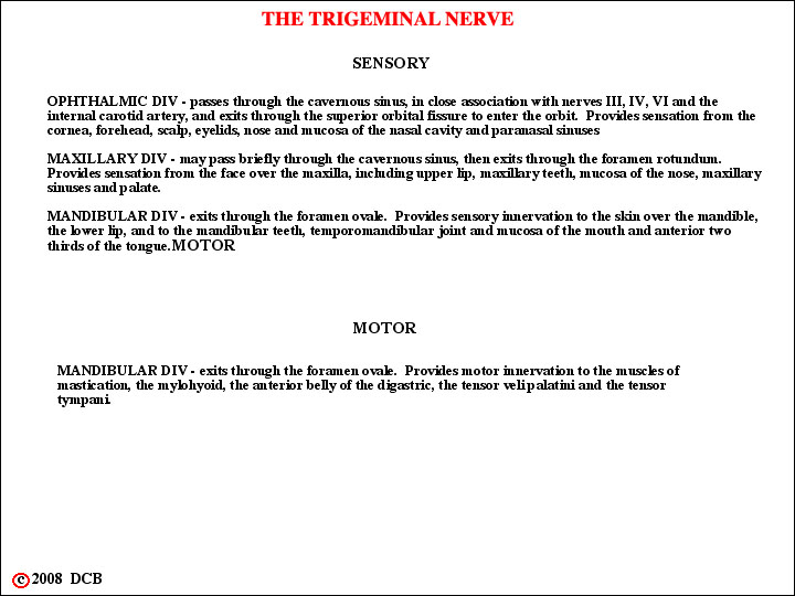
THE TRIGEMINAL NERVE
This figure and the next have the details.
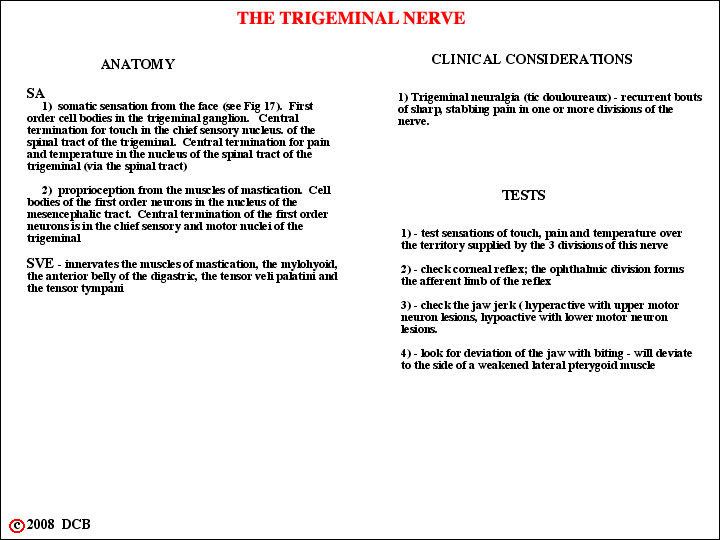
THE TRIGEMINAL NERVE
This figure and the previous have the details.
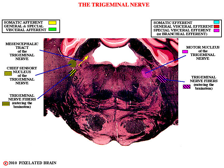
THE TRIGEMINAL NERVE
This shows the nucleus that gives rise to the motor component of the nerve and the chief sensory nucleus. We will see the other sensory nuclei when we go through the slide series.