MODULE 9 - VIEWS-ALL
The
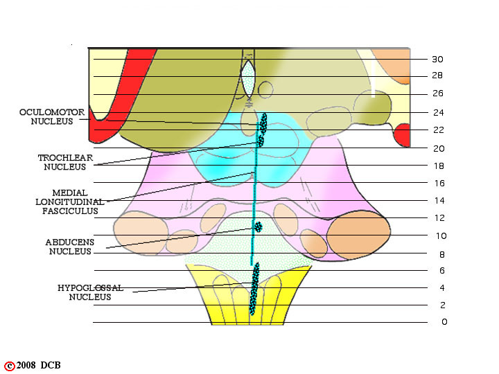
FIGURE 9-1
1

FIGURE 9-2
The trochlear nerve is unusual in that its axons, after leaving the trochlear nucleus (present in the midbrain), arch caudally and dorsally around the edge of the periaqueductal gray, decussate in the anterior medullary velum and emerge from the brainstem on its dorsal surface at the junction of the pons and midbrain just caudal to the inferior colliculus.
The trochlear nucleus is seen only in Slide 21 . The exiting fibers pass caudally and "wrap around" the cerebral aqueduct in Slide 20, Slide 19, Slide 18 and Slide 17. They then decussate in the anterior medullary velum and depart from the brain in Slide 16.
26 27 21 22 23 24 25 16 17 18 19 20
2
FIGURE
z
FIGURE
z