MODULE 10 - SECTION 3 - THE POSTERIOR ARTERIAL SUPPLY to the BRAIN
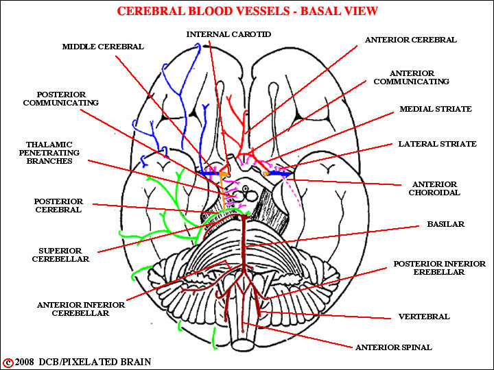
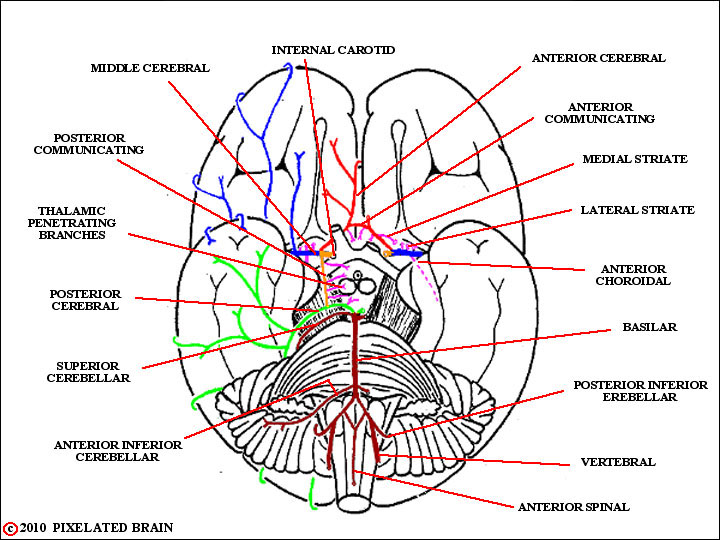
- - - This part of the system is fed by the two vertebral arteries which join at the border between the medulla and pons to form the basilar artery(Figure 10-3 again). This is the only example we can think of in the body in which two arteries join as they pass to the periphery to form a single, larger vessel - and one can't help but wonder if the "purpose" is to insure a constant blood supply to that relatively small part of the brain (the brainstem) the function of which is essential for the maintenance of life.
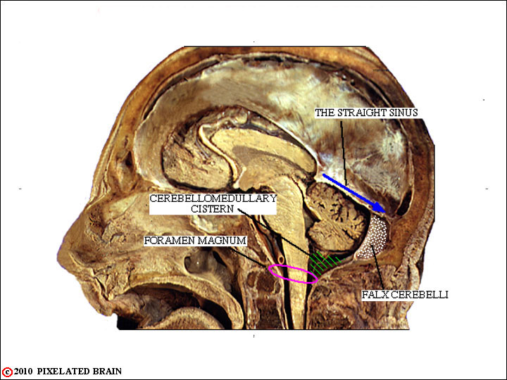
enters through the
FORAMEN MAGNUM
In the lower part of this figure the left vertebral has been cut just below the point where it joins the right vertebral to form the basilar artery; just above this, the basilar can be seen running between the basilar pons and the base of the skull.
BLOOD SUPPLY of the BRAIN
a BASAL VIEW
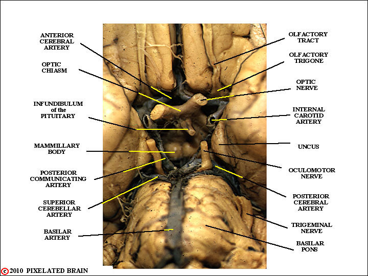
The basilar artery ends by giving off two branches, the superior cerebellar and posterior cerebral arteries . The oculomotor nerve passes between these two vessels as it exits from the brainstem; this is an important relationship.
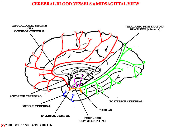
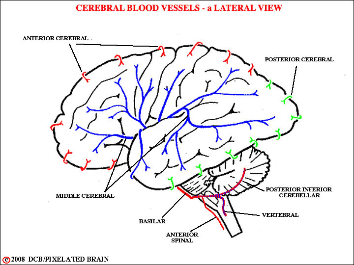
The posterior cerebral artery supplies the medial surface of the occipital lobe and the basal surface of the occipital and temporal lobes. In addition, like the anterior cerebral, it sends branches around the "corner" on to the lateral surface of the hemisphere.
Look at the next 3 slide sections for the blood supply to the hemisphere and the maps given in Blumenfeld's Figures 10.5 & 10.9 to confirm the region of the hemisphere supplied by the posterior cerebral artery.
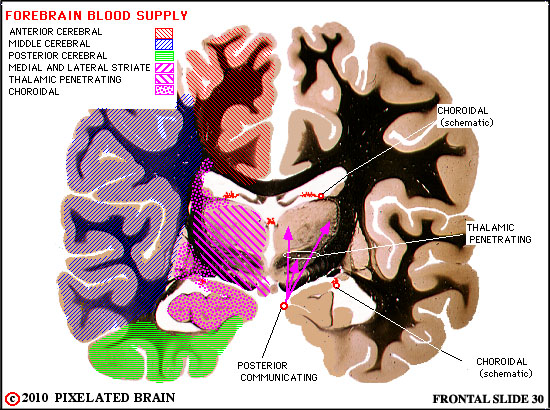
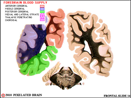
FOREBRAIN BLOOD SUPPLY
SLIDE 30
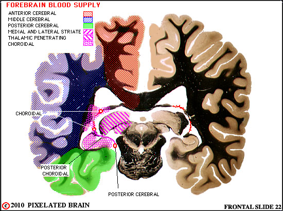
FOREBRAIN BLOOD SUPPLY
SLIDE 22
FOREBRAIN BLOOD SUPPLY
SLIDE 14
 BLOOD SUPPLY of the BRAIN
a BASAL VIEW
BLOOD SUPPLY of the BRAIN
a BASAL VIEW
 THE CIRCLE of WILLIS
and
SURROUNDING AREA
THE CIRCLE of WILLIS
and
SURROUNDING AREA
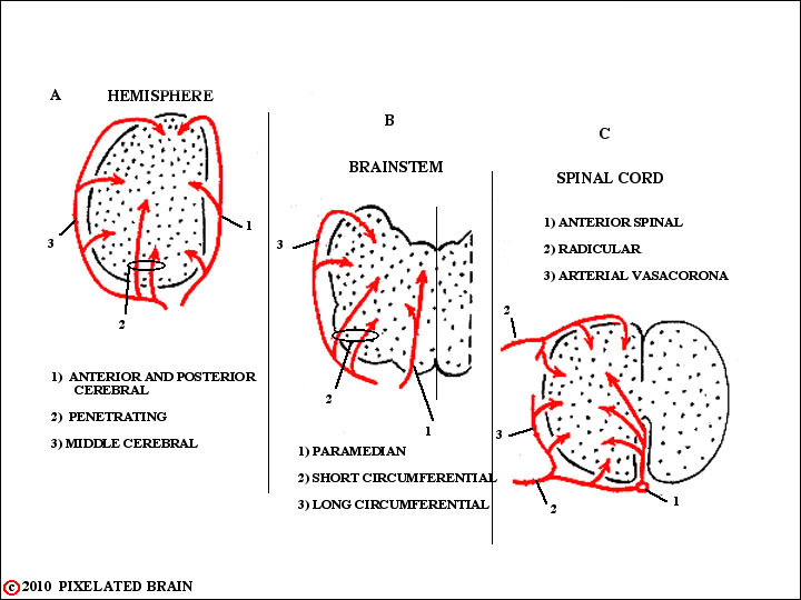
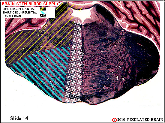
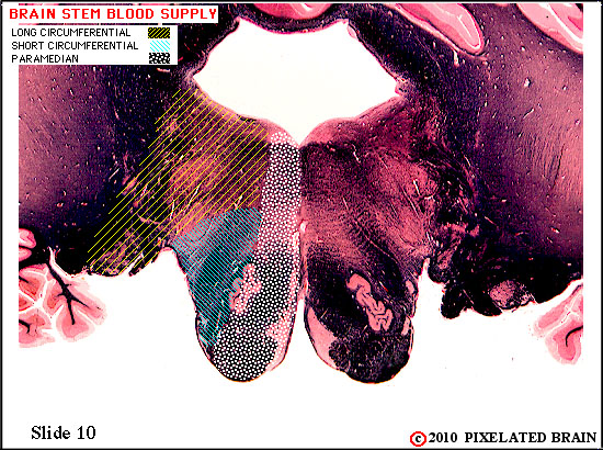
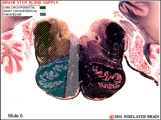
THE PATTERN of BLOOD SUPPLY
is the SAME at all levels of the
CENTRAL NERVOUS SYSTEM
The basic plan of the blood supply to the brain stem is the same at all levels and is illustrated here. The long circumferential branches are derived, in ascending order, from the posterior inferior cerebellar artery, the anterior inferior cerebellar artery, the superior cerebellar artery and the posterior cerebral artery.
Now look at our sections for the blood supply to the brainstem.

BRAINSTEM BLOOD SUPPLY
SLIDE 18
BRAINSTEM BLOOD SUPPLY
SLIDE 14
BRAINSTEM BLOOD SUPPLY
SLIDE 10
BRAINSTEM BLOOD SUPPLY
SLIDE 6