MODULE 10 - SECTION 5 - BLOOD SUPPLY of the SPINAL CORD

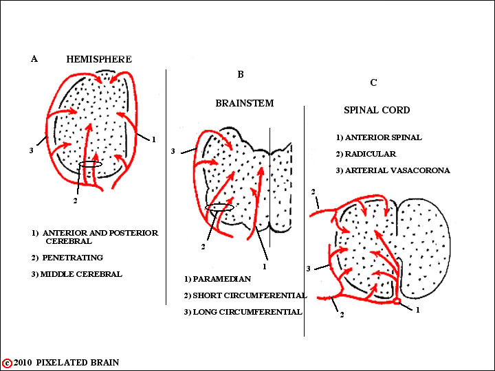
THE PATTERN of BLOOD SUPPLY
is the SAME at all levels of the
CENTRAL NERVOUS SYSTEM
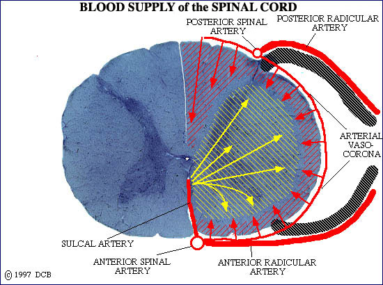
- - - If you look at this figure and consider what you have seen so far, you will predict that one group of blood vessels will pass over the surface of the spinal cord, sending blood to the zone immediately within, while other branches (penetrating vessels) will supply the central region of the cord. Congratulations!, you are right. Posterior intercostal arteries give off segmental branches which enter the vertebral canal through intervertebral foramina, join spinal nerves and then divide into anterior and posterior radicular arteries. The radicular arteries run medially on dorsal and ventral roots to reach the cord. Once there, they do two things
----- 1) Branches pass over the surface of the cord and supply the tissue immediately deep to the surface. These are termed the arterial vasocorona (Cu Slide C6) or the spinal arterial plexus.
----- 2) In addition, the radicular arteries contribute to longitudinal vessels, the anterior and posterior spinal arteries. The anterior spinal artery, in turn, sends sulcul branches into the ventromedian sulcus (labeled ventromedian fissure in Figure 2-31) to supply the interior of the cord. Oddly enough, these sulcul branches turn alternately to the right and left, each supplying only half the cord. While the anterior spinal artery is truly a single vessel as depicted on Cu Slide C6, the paired posterior spinal arteries are each actually a plexus of vessels with many connections between the small vessels.
 Slide C-6
- - - There is one important detail to add to this description. While textbooks vary in the way they put it, the fact is that not all spinal arteries send branches to the cord in the manner illustrated on this slide (C-6). Rather, the contribution to the anterior and posterior spinal arteries is much more limited. Blood running up and down the cord in the anterior and posterior spinal arteries supplies the segments that do not have radicular arteries of their own. While the posterior spinal artery easily does the job because of the many anastomotic connections that characterize it, the anterior spinal artery is more variable, and thus heavily dependent on the integrity of the radicular arteries that feed it. If one of the posterior intercostal vessels feeding a key artery (the great artery of Adamkiewicz, for example) is inadvertently clamped during surgery, severe spinal cord damage of the territory supplied by the anterior spinal artery may result. Checking this slide (C-6) again, it will be apparent that if the anterior spinal artery is deprived of blood, the anterior 2/3 of the cord, including all descending motor pathways, will be damaged, but the ascending pathways in the dorsal columns will remain intact.