MODULE 11 - VIEWS-ALL
The
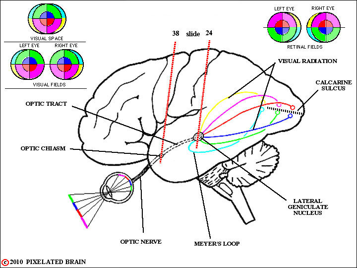
FIGURE 11-1
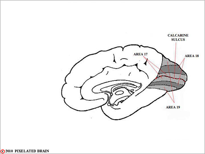
Note that information coming from the upper part of visual space (the green and dark blue bars in the visual target in the lower left part of the view) falls on the lower part of the retina, and ultimately projects to the lingula, the gyrus inferior to the Calcarine Sulcus. Conversely, information from the lower part of visual space (the purple and red bars in the targe) falls on the upper part of the retina, and ultimately projects to the cuneus,the gyrus superior to the Calcarine Sulcus.
The red and dark blue colors are intended to represent the pathway conveying information from the central part of the retina - the macula - to the visual cortex. Note that these pathways terminate most posteriorly in the cortex. The green and purple colors represent the pathway conveying information from the peripheral part of the retina. Note that these pathways terminate more anteriorly in the cortex.
The yellow and light blue colors are best ignored in this view.
THE VISUAL PATHWAY
We will organize our description of the visual system into six parts, which correspond to six regions of the visual pathway.
1) The region anterior to the optic chiasm.
2) The optic chiasm.
3) The region between the optic chiasm and the lateral geniculate nucleus - in other words, the region where the optic tract conveys information centrally.
4) The level of the thalamic relay for visual information - through the lateral geniculate nucleus.
5) The pathway by which visual information traverses the internal capsule to reach the visual cortex - termed the visual radiation.
6) The visual cortex.

FIGURE 11-2
The reason to look at this view now is to be reminded of the intimate relationship between the optic chiasm and the Circle of Willis. Perhaps the most important relationship is the one between the internal carotid artery and the lateral edge of the chiasm, but note also that the anterior cerebral arteries run medially just above the "incoming" optic nerves. You can imagine a variety of visual fields deficits that could result if these vessels were to press on the chiasm. The infundibulum of the pituitary should remind you that the pituitary is lurking just underneath the chiasm and also capable of causing trouble.
FIGURE 11-3
40 __38 __36 __34 __32 __30 __28 __26 __25 __24a __24b __22 __20
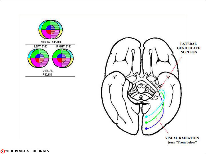
This view simply gives you a way of calling up slides and keeping track of where you are. You may, however, find it easier to start by clicking on Slide 40 and then work your way through the series using the "caudal" buttons. Note that while the optic tract may appear to be "inside" the brain, it actually just runs laterally over the surface of the cerebral peduncle to reach the lateral geniculate nucleus. The basal view of the brain, diganat_1h.jpg , shows the tract starting to move laterally, and the brain dissection, diganat_2q.jpg , traces it all the way to the lateral geniculate nucleus .
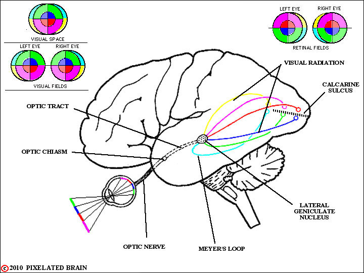
FIGURE 11-4
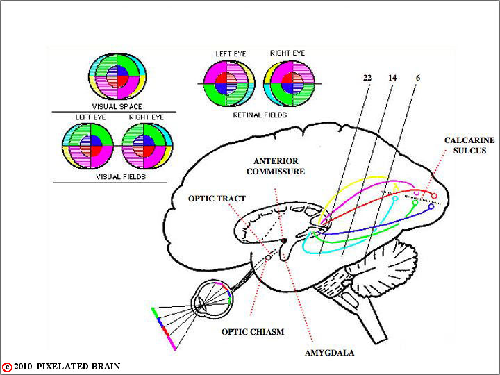
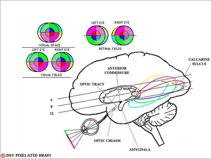
This view tries to show the relationship between visual space, the visual fields, the retinal fields (our term) and the anatomy of the visual radiation.
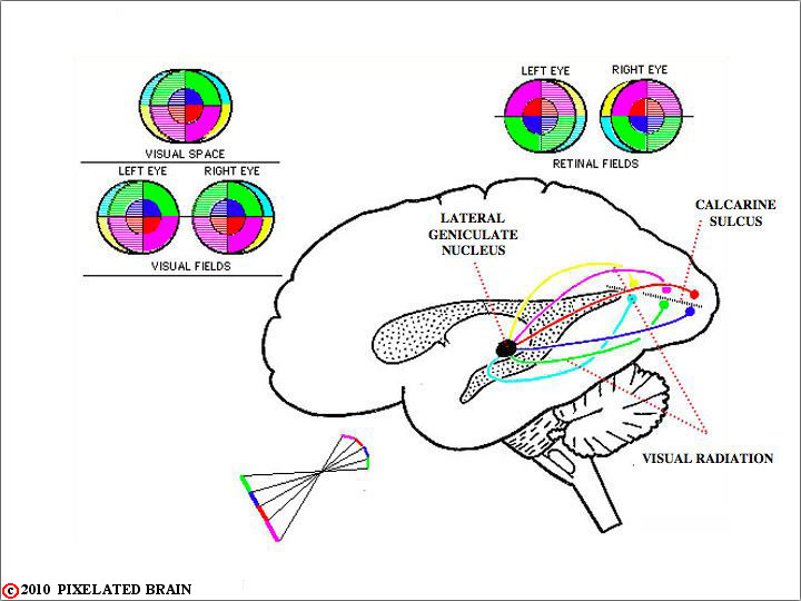
FIGURE 11-5
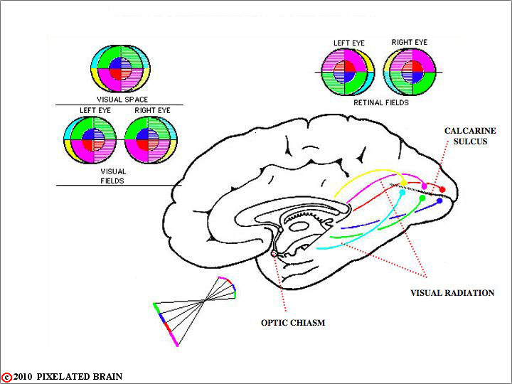
FIGURE 11-6