MODULE 11 - SECTION 7 - THE VISUAL RADIATION
In the preceeding section we showed the retinotopic organization of the first part of the visual pathway primarily for the sake of completeness. It is only at the level of the chiasm that the precise course of the fibers is important. Elsewhere - in the optic tract, for example - the fibers are packed together so closely that a lesion that gets some of them is likely to get them all. Thus the exact spatial relationship between fibers from different parts of the retina probably doesn't matter.
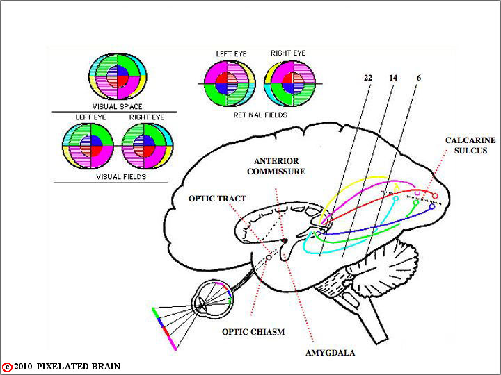
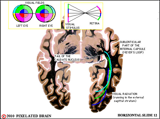
However, as the pathway leaves the lateral geniculate nucleus, and heads for the visual cortex within the visual radiation, things change. The fibers fan out in such a way that there is a very good chance that some may be damaged while others remain intact. When this happens certain characteristic visual field deficits are produced. These are discussed in Blumenfeld starting on Page 442. The purpose of the figures included in this section is to help you visualize this part of the pathway.
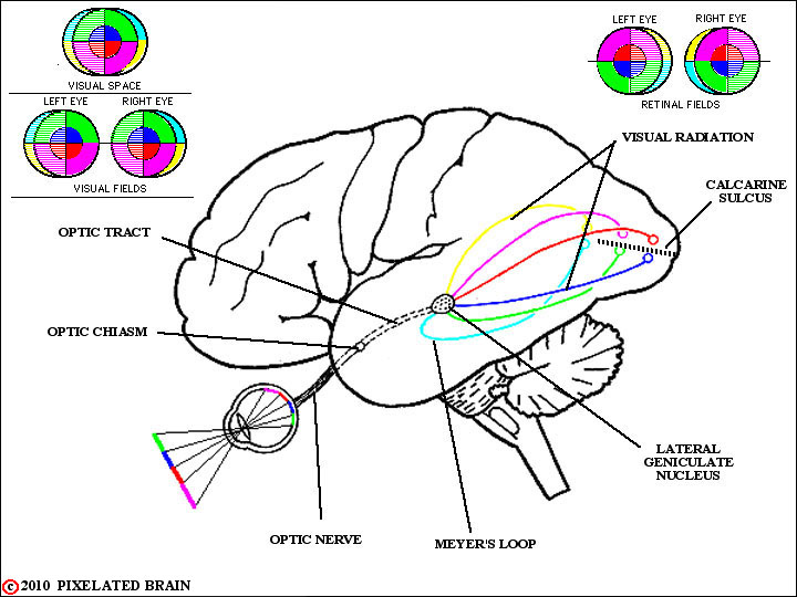
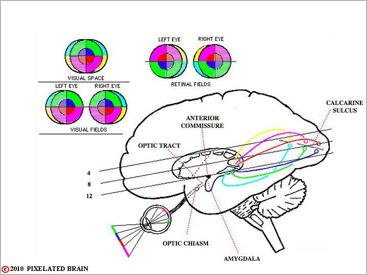
THE VISUAL RADIATION on a LATERAL VIEW of the BRAIN
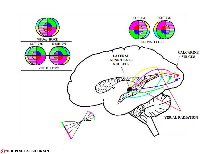
VISUAL RADIATION to the lateral ventricle SHOWN on a lateral view
of the BRAIN
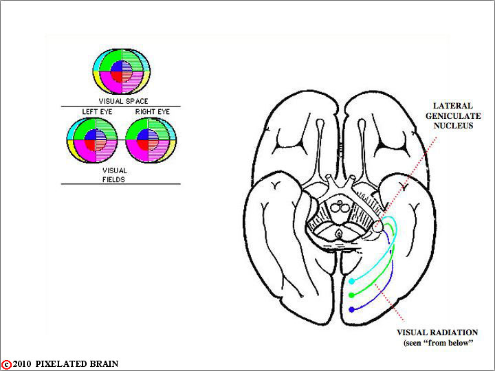
BASAL VIEW of the BRAIN
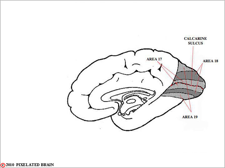
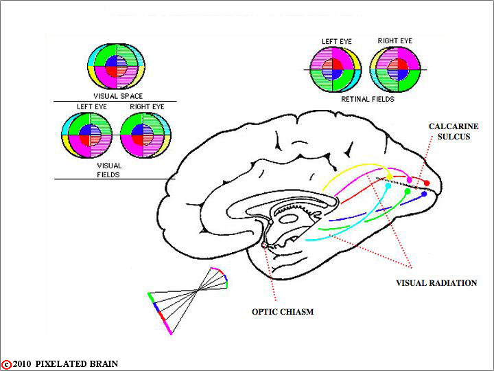
MEDIAL VIEW of the BRAIN

THE VISUAL PATHWAY on SLIDE 22
XX

THE VISUAL PATHWAY on SLIDE 14
XX
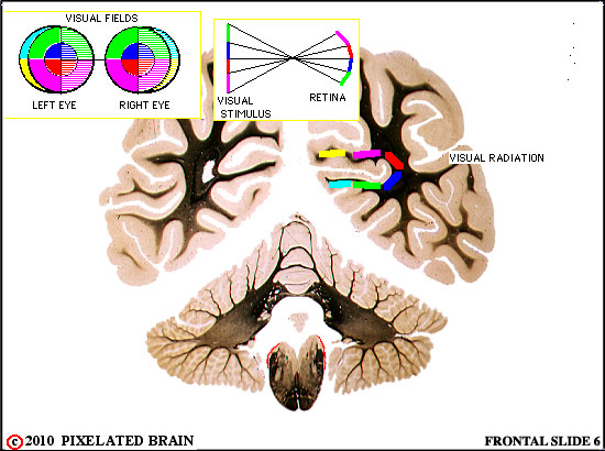
THE VISUAL PATHWAY on SLIDE6
XX

THE VISUAL PATHWAY on SLIDE4
XX
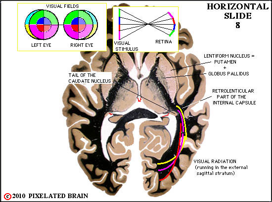
THE VISUAL PATHWAY on SLIDE8
XX
XX
XX