MODULE 12 - SECTION 1 - THE EXTERNAL. MIDDLE and INNER EARS
HOW THE AUDITORY SYSTEM IS STIMULATED
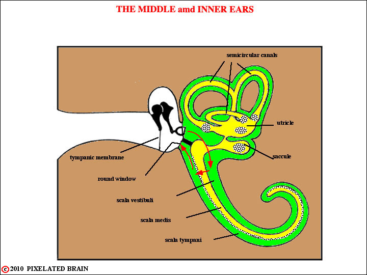
Let's start with a simplified version of this remarkable system and then come back to consider parts of it in greater detail. Variations in air pressure (i.e. sound) pass through the external acoustic meatus and medially in the ear canal to impinge upon the tympanic membrane . Movement of the tympanic membrane is transmitted by the bones of the middle ear to the foot plate of the stapes, positioned within the oval window of the inner ear . This in turn causes deflections of the basilar membrane, which travel from the base to the apex of this structure. The basilar membrane is "tuned" in such a way that low frequencies resonate near the apex and high frequencies resonate near the base. When the basilar membrane moves, the hair cells that lie on it are alternately hyperpolarized and depolarized ; depolarization initiates action potentials in the peripheral terminals of the first order neurons of the auditory pathway. These neurons have their cell bodies in the spiral ganglion , buried within the modiolus , and send their central processes through the internal acoustic meatus to enter the cranial cavity.
We strongly encourage you to look at some excellent animations of the events described above. To do so click on ear animation and look at the topics listed below - if possible, view these animations using Safari:
- Middle ear - you have to click on the left triangle repeatedly to view it. Note how the round window membrane bulges out when the stapes moves in
- Ossicle Motion - shows how the ossicles move about a pivot axis
- Cochlear Motion - shows how everything moves as a pressure wave travels up the the scala vestibuli
- Corti Motion - shows how the stereocila of the outer hair cells touch the tectorial membrane and wiggle as the basilar membrane and organ of corti move. See Haines Figure 21-4 on Page 338 for comparison.
- Corti Motion - shows how the stereocilia of the inner hair cells move, too, even though not in contact with the tectorial membrane
- HC Excitation - shows that when the short stereocilia are bent towards the tallest one the hair cell is depolarized and first order afferent fibers fire.
- HC Inhibition - shows the reverse of the above
- Traveling Waves - shows how deflections travel up the basilar membrane when 6 different frequencies of sound (top left =500 cps, bottom right = 16,000 cps) are played into the ear. Note that the region of maximum deflection is near the helicotrema for low frequencies and near the base for high frequencies. Compare with Haines Figure 21-2 on Page 336 for comparison
Things are, however, more complex than they seem because it has been clear for years that the mechanical tuning of the basilar membrane is rather broad - particularly for low frequencies - and cannot, by itself, account for the way in which sound waves are so precisely translated into a neural code.
HOW THE VESTIBULAR SYSTEM IS STIMULATED
The vestibular system detects the fixed position of the head in space, and both linear and angular acceleration. Hair cells in the macula of the saccule and utricle accomplish the first of these tasks. The hairs of these hair cells are imbedded a gelatinous structure containing crystals of calcium carbonate. If the head is held in a tilted position, the weight of the crystals will, via the gelatinous layer, bend the hairs of the hair cells. Because of the physical orientation of the hair cells in the saccule and utricle, any given head position will result in a unique pattern of neural activity which allows the brain to determine what the position is. These same organs (the saccule and utricle) can detect linear acceleration - partly accounting for the sensation you experience when you sit in a car and floor the gas pedal. In contrast, the semicircular canals are involved in the detection of angular acceleration. When the head suddenly turns, for example, the endolymph in one or more canals lags behind. The resultant activation of a selective population of hair cells advises the brain about the details of the acceleration.
1
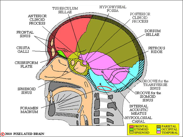
INTERIOR of the SKULL
The vestibulocochlear nerve enters the cranial cavity by way of the internal acoustic meatus.
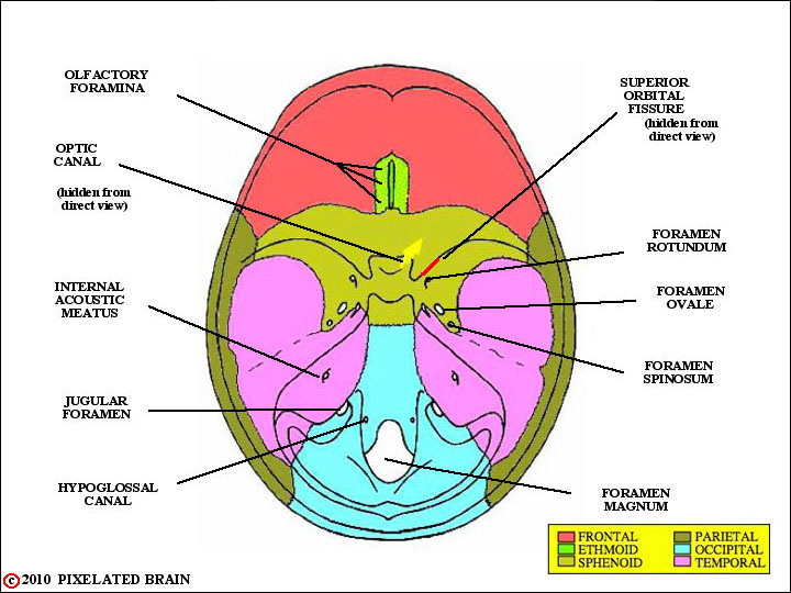
FORAMINA in the SKULL
a BASAL VIEW
The vestibulocochlear nerve attaches to the brain in a region called the cerebellopontine angle. This region lies just caudal to the basilar pons and just dorsolateral to the rostral pole of the inferior olive. The flocculus and ventral edge of the cerebellum are nearby. The facial nerve, which also enters the cranial cavity through the internal acoustic meatus, is just medial to the vestibulocochlear one . Thus, in lesions of the cerebellopontine angle both nerves are often damaged.