MODULE 12 - SECTION 5
FLASH CARDS for the AUDITORY and VESTIBULAR SYSTEMS
In this computer version of flash cards, questions alternate with answers as you scroll down the page. We suggest that you limit the height of your viewing window so that you can't see the answers until you have made your own choices. In this first exercise, we just ask you to identify structures or spaces. In future sets of flash cards we expect to use the blue boxes on the right to ask functional questions.
We will be adding "answers" for these cards shortly.
QUESTION 1
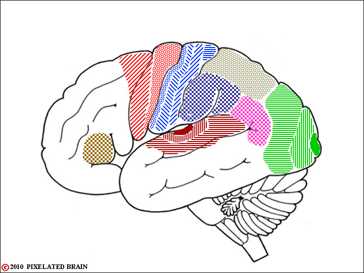
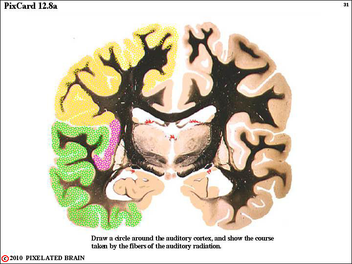
The auditory cortex is axtually hidden from view, in Brodmann's cortical area 41. Auditory information is passed from there to an adjacent cortical area named Wernicke's area (area 22), where the initial processing of language takes place. This area has reciprocal connections with the Angular gyrus (area 39) and the Supramarginal Gyrus (area 40) which share in this function.
We have used shading to outline all these cortical areas. Try to identify each of them by adding the appropriate Brodmann numbers.
Next identify Broca's area, which lies in the pars triangularis (area 45) of the inferior frontal gyrus.
Finally, draw in the arcuate fasciculus, which connects Wernicke's area with Broca's.
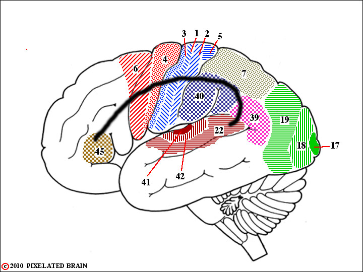
ANSWER 1
QUESTION 2
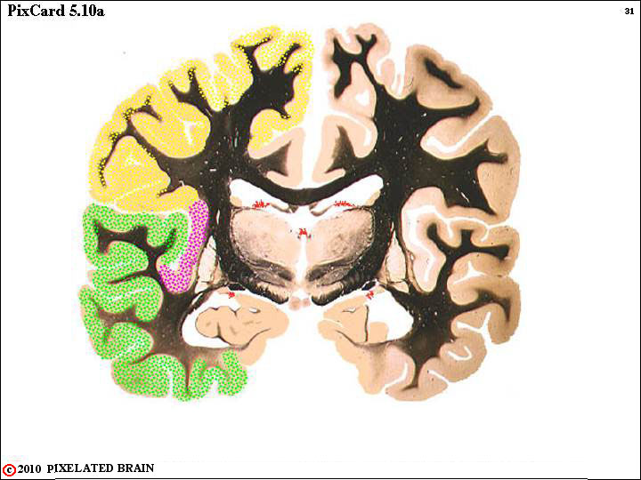
Draw in the auditory radiation, showing how it passes through the sublenticular part of the internal capsule to reach the temporal lobe.
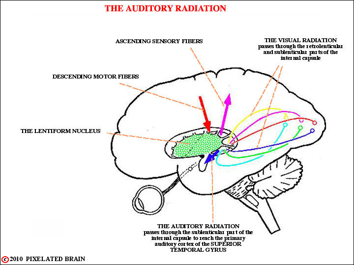
ANSWER 2
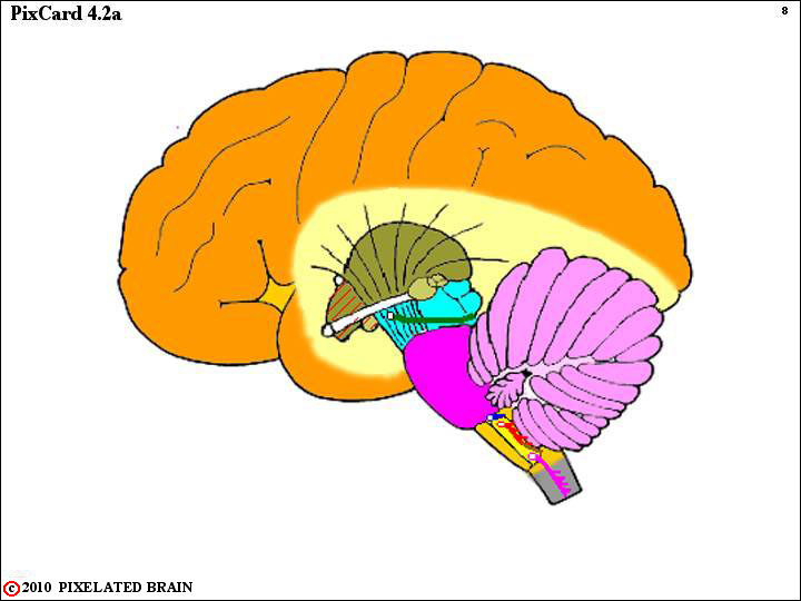
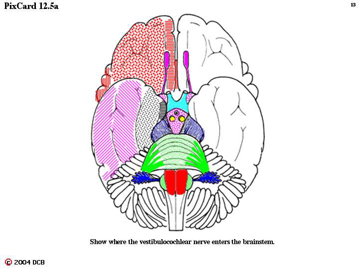
QUESTION 3
Label the inferior colliculus and the medial geniculate nucleus.
Next, draw in and label the pathway that runs between these two structures.
Finally crrcle and label the region of the cerebellopontine angle.
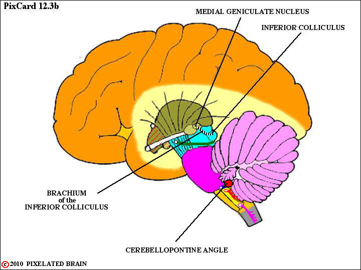
ANSWER 3
z
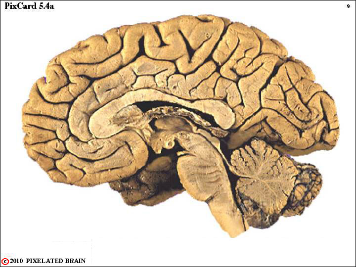
QUESTION 4
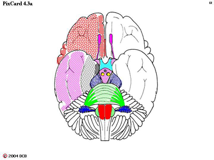
Circle the inferior colliculus.
In what division of the brainstem is this structure found? ___________ Use lines to define the rostral and caudal borders of this division.
Which two cranial nerves exit the brainstem at this level??
________________________
________________________
One of these exiting nerves can be seen in this view, Label it.
The second nerve cannot be seen. Mark with an "X" the region where this nerve will exit.
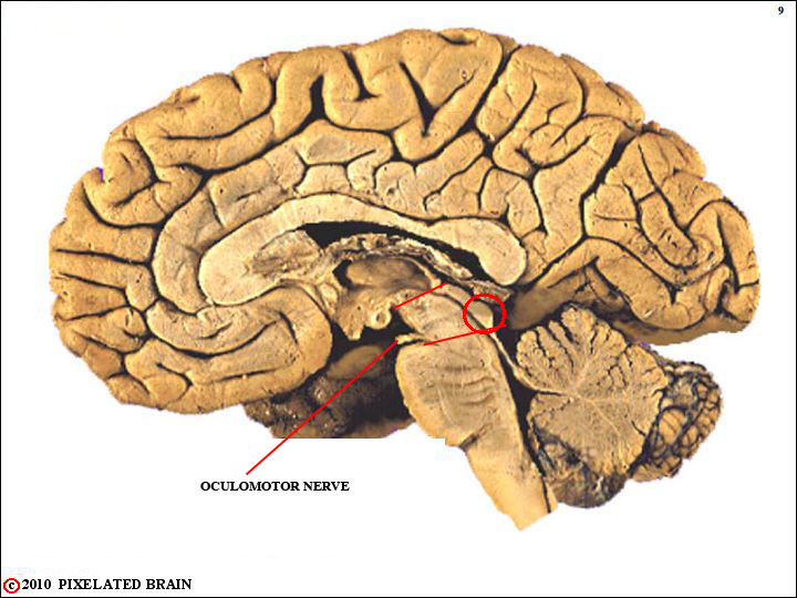
ANSWER 4
QUESTION 5
ANSWER 5
QUESTION 6
ANSWER 6
QUESTION 7
ANSWER 7
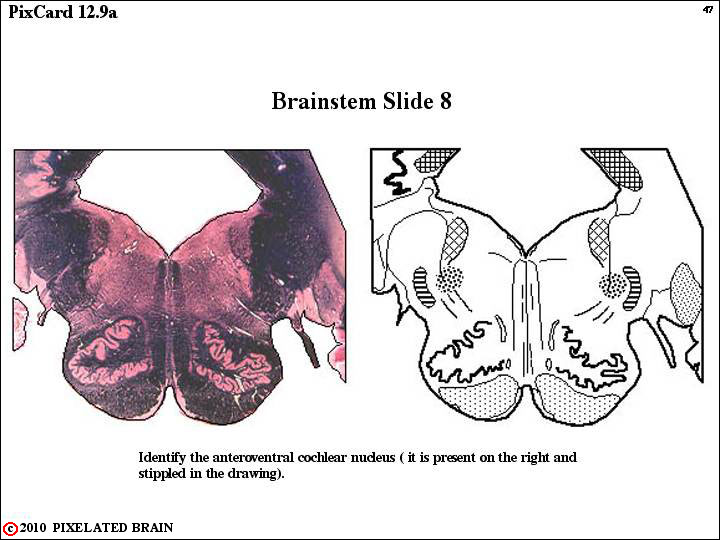
QUESTION 8
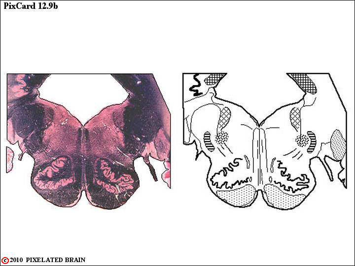
ANSWER 8
QUESTION 9
ANSWER 9
QUESTION 10
ANSWER 10
QUESTION 11
ANSWER 11
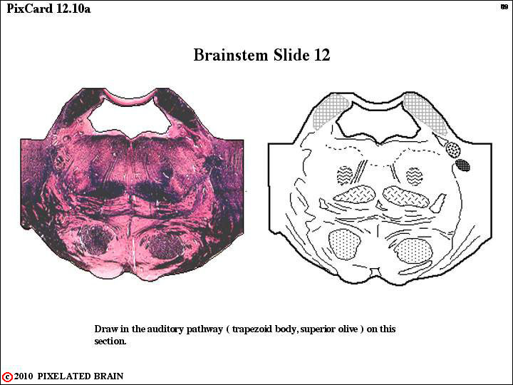
QUESTION 12
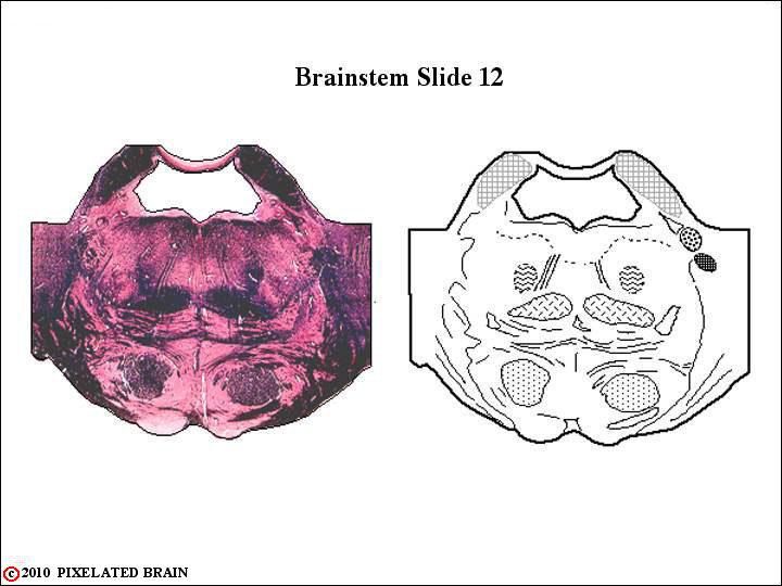
ANSWER 12
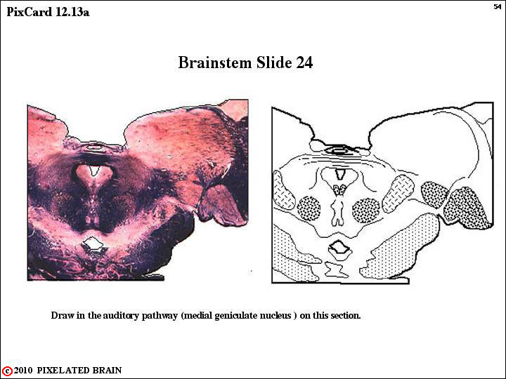
QUESTION 13
ANSWER 13
QUESTION 14
ANSWER 14