MODULE 13 - SECTION 5 - THE HYPOTHALAMUS
The position of the hypothalamus within the brain is best seen in mid-sagittal views. The first one relates the hypothalamus to other regions, and the next defines the borders of the diencephalon (of which the hypothalamus is a part). The following view reminds you that the third ventricle separates the region into two halves and the last one shows how the hypothalamic sulcus forms a border between the thalamus and the hypothalamus. Our aim in these first few slides is simply to make certain you know where the hypothalamus is.
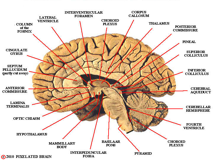
LANDMARKS
on a MIDSAGITTAL VIEW
of the GROSS BRAIN

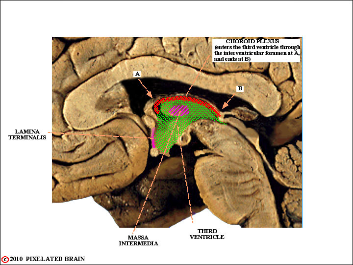
LAMINA TERMINALIS,
CHOROID PLEXUS and the
third ventricle
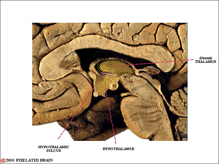
THE HYPOTHALAMIC SULCUS
This last view shows how the hypothalamic sulcus forms a border between the thalamus and the hypothalamus.
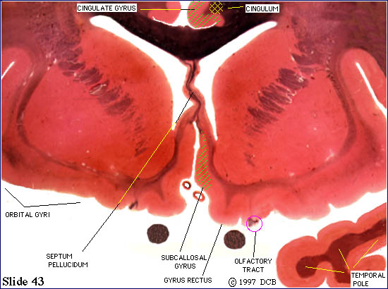
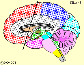
SLIDE 43
Anterior to the Septal Level
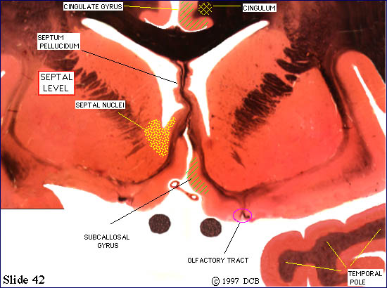

SLIDE 42
Anterior to the Septal Level
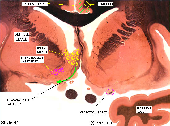
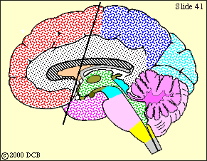
SLIDE 41
Septal Level
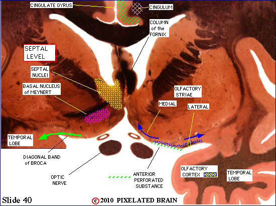
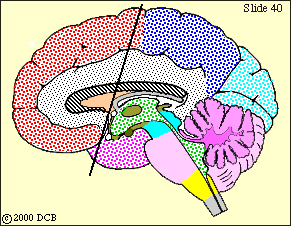
SLIDE 40
Septal Level
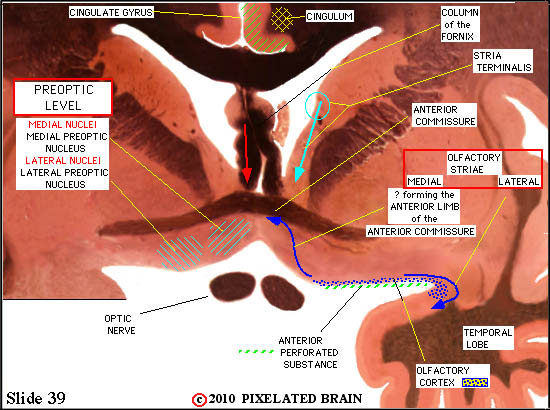
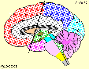
SLIDE 39
Preoptic Level
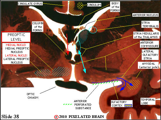
SLIDE 38
Preoptic Level
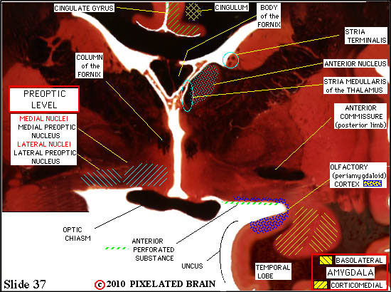

SLIDE 37
Preoptic Level
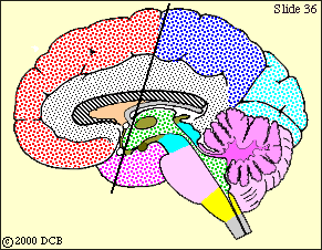
SLIDE 36
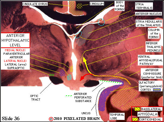
Anterior Hypothalamus
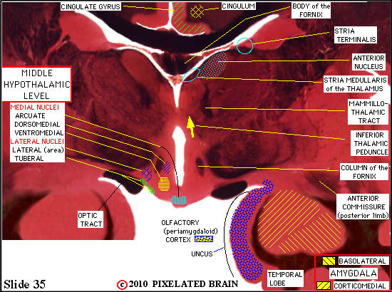
SLIDE 35
Middle Hypothalamic Level
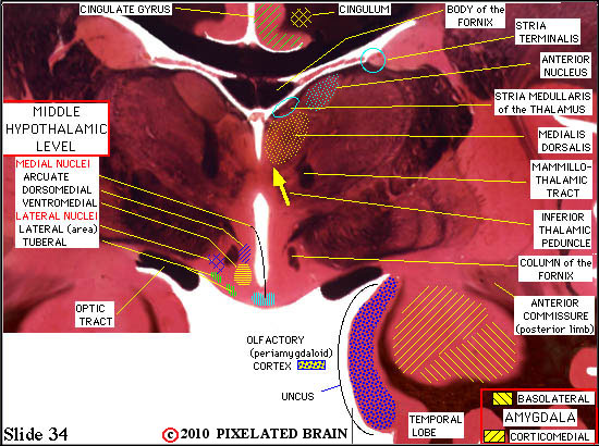

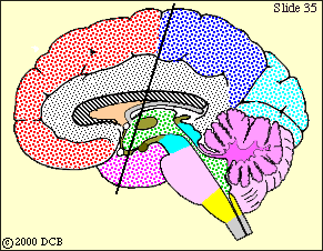
SLIDE 34
Middle Hypothalamic Level
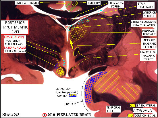

SLIDE 33
Posterior Hypothalamic Level
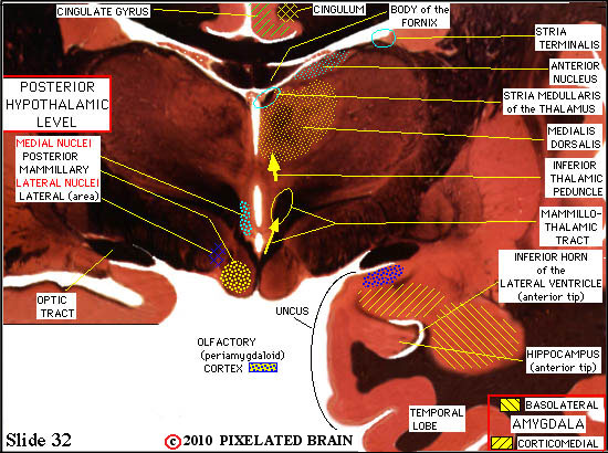

SLIDE 32
Posterior Hypothalamic Lev
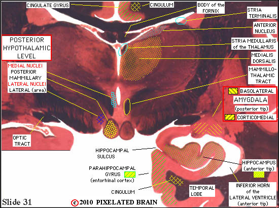
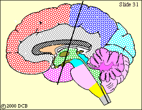
SLIDE 31
Posterior Hypothalamic Level
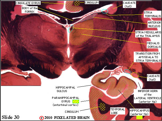
SLIDE 30
Hippocampal Formation