MODULE 14 - SECTION 2 - THE FOUR SUBDIVISIONS of the DIENCEPHALON

DIVISIONS of the BRAINSTEM
You already know that there are four subdivisions of this region.
of the
GROSS BRAIN - QUIZ VERSION
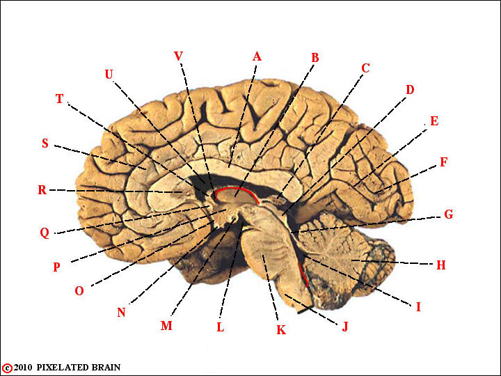
The relative positions of these regions is best seen in sagittal views of the brain. Look first at this midsagittal view of the gross brain. Just for fun, try this unlabelled version first and impress those around you by rattling off the names of all the structures marked. Then, if you have any doubts, look at the labeled version on the next slide.
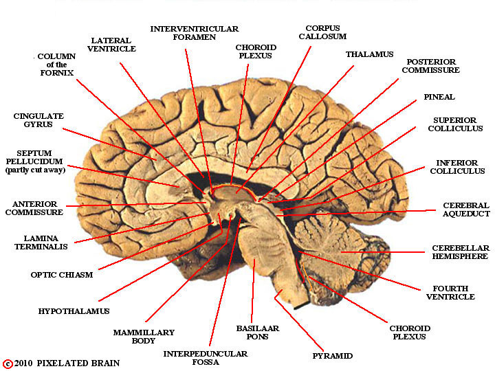
on a MIDSAGITTAL VIEW
of the GROSS BRAIN

BORDERS of the DIENCEPHALON
A plane (A) passing just rostral to the superior colliculus dorsally and just caudal to the mammillary bodies ventrally separated the diencephalon from the midbrain. A second plane (B) passing through the anterior commissure and the optic chiasm marks the rostral or anterior limit of the diencephalon (and of the primitive neural tube).
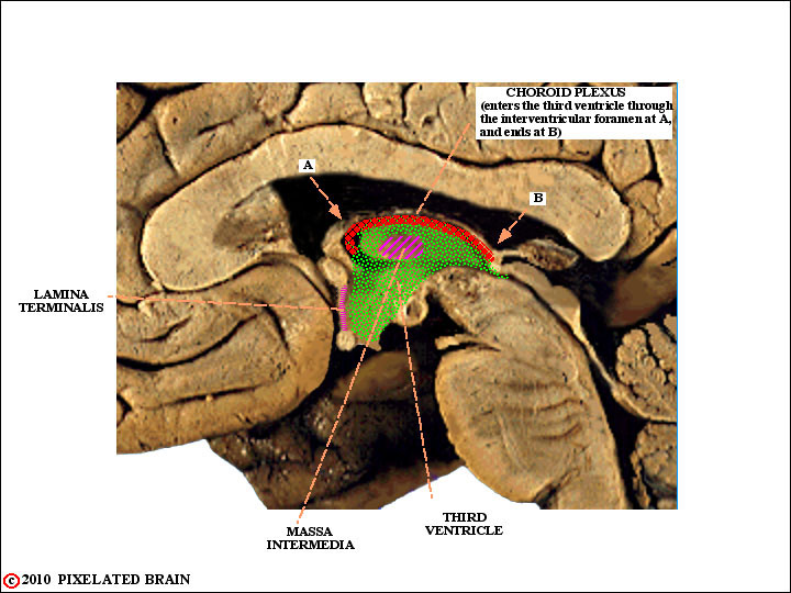
THE THIRD VENTRICLE
and the
CHOROID PLEXUS
Except for a limited region, the massa intermedia (which is present in most but not all brains) the diencephalon on one side is separated from that on the other side by the third ventricle. The third ventricle is limited dorsally by choroid plexus and anteriorly by the lamina terminalis.
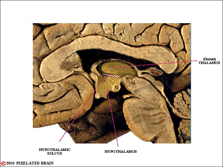
THALAMUS, HYPOTHALAMUS
and the
HYPOTHALAMIC SULCUS
The hypothalamic sulcus, a small recess in the lateral wall of the third ventricle, serves to define the border between the two largest subdivisions of the diencephalon - the thalamus and the hypothalamus.
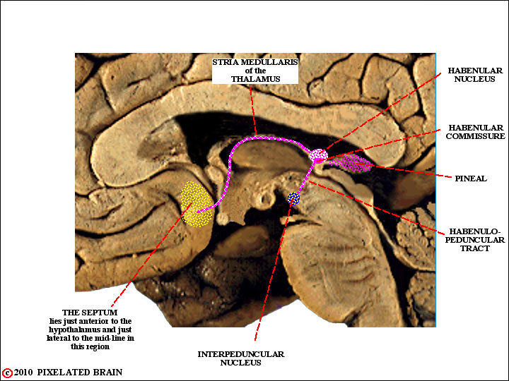
THE EPITHALAMUS
and
RELATED STRUCTURES
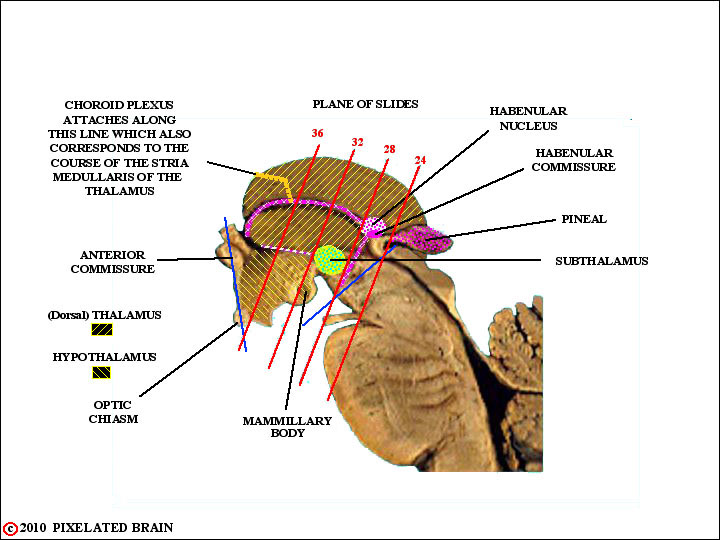
The third diencephalic region - the epithalamus - consists of several structures which have been drawn in on this view. The habenular nuclei and associated pathways are considered a part of the limbic system but we know very little about their function.
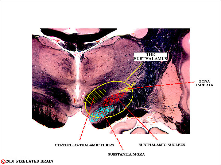
THE SUBTHALAMUS
The final subdivision of the diencephalon is the subthalamus. This limited region lies ventral to the thalamus at the juncture between the diencephalon and the midbrain, and is best seen in frontal views such as this one. From our point of view, the most significant structure in this region is the subthalamic nucleus and we consider it in detail when we deal with the basal ganglia.
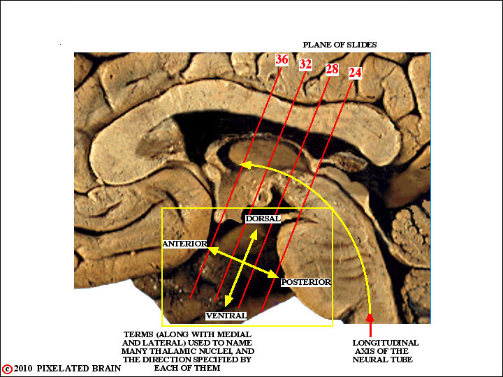
PLANES and DIRECTIONS
in the
DIENCEPHALON
Because of the bend in the longitudinal axis of the neural tube at the midbrain level, our slides pass through the diencephalon in a true frontal plane. That's a break, because it means that they coincide with the orientation of the dorsal-ventral, anterior-posterior scheme used to name thalamic nuclei. Looking at this view alone, you might conclude that the thalamus is not much bigger than the hypothalamus and that neither extends caudally as far as slide 25.

THE DIENCEPHALON
You would be wrong on both counts. Lateral to the midline, the thalamus expands dorsally to become by far the larger structure.
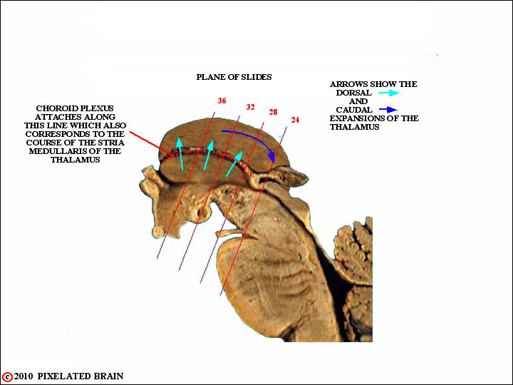
THE DORSAL THALAMUS
extends caudally over the
MIDBRAIN
The dorsal thalamus also extends caudally to cover part of the midbrain, and first appears in slide 19. Thus, a midsagittal view in which interfering structures have been cut away would look like this.
THE DIENCEPHALON on SLIDES
The last view of this series summarizes many of the relationships that have been described.