MODULE 12 - SECTION 2 - THE AUDITORY PATHWAY
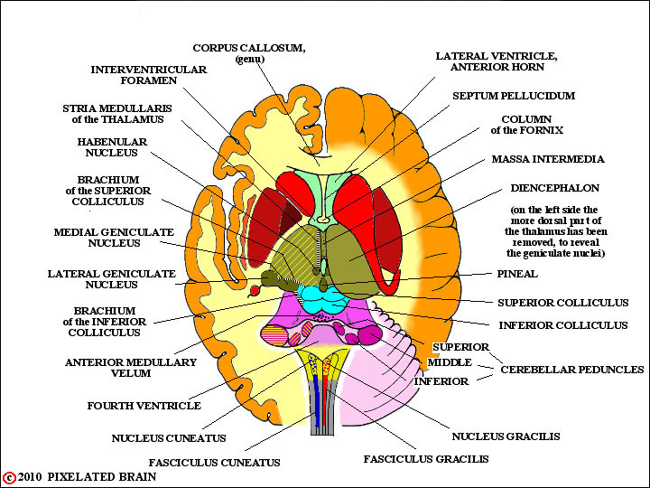
DORSAL VIEW of the BRAINSTEM
A good way to start your review of auditory pathways is by looking at it, drawn on a dorsal view of the brain such as the figure here.
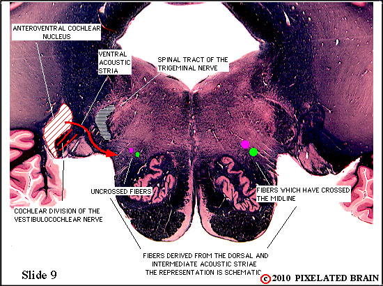
AUDITORY PATHWAY on SLIDE 9
- - - The cochlear division of vestibulocochlear nerve enters the brain at the level of Cornell Slide 9, and the two red lines in this view show the position and course of the fibers. Most entering first order neurons give off two collateral branches; one of these terminates in the ventral cochlear nucleus and the other passes dorsally over the inferior cerebellar peduncle to end in the dorsal cochlear nucleus. This nucleus lies on the dorsal surface of the inferior cerebellar peduncle, just lateral to the inferior vestibular nucleus. This arrangement is shown schematically in Haines' Figure 21-9. This is a good example of divergence and suggests that auditory information passes rostrally in the brain by way of two parallel pathways - each coded for a different parameter of information. This arrangement should remind you of the M and P pathways in the visual system.
Use this link to see annotated slides:
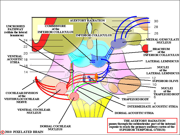
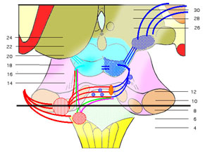
AUDITORY PATHWAY SLIDE LEVELS on a DORSAL VIEW of the BRAINSTEM