PIXBRAIN HOME _ _ MOD 1 HOME _ _ previous _ _ FIGURE 1-41 _ _ next _ _ I WANT TO
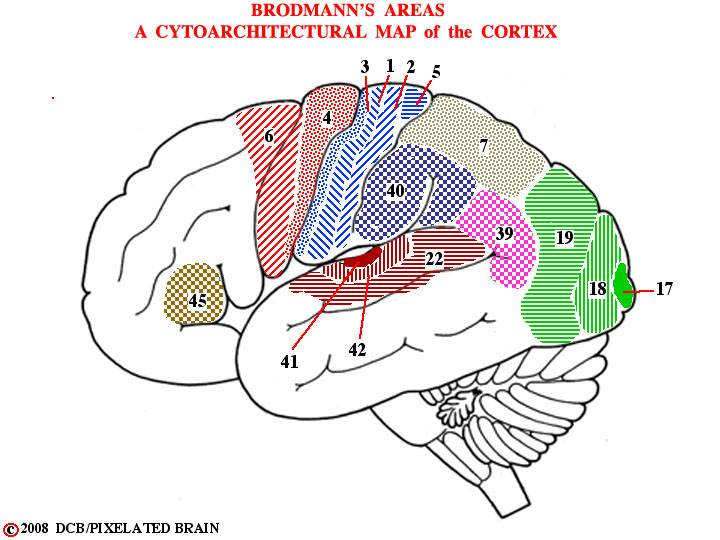
- - At this point, you are asking "Is that it? Do I have all the names I need to describe the surface of the cerebral cortex? If so, now tell me what each of these gyri actually does." Well, the gyri are a necessary start, but its not that simple (it never is). The gyri are useful landmarks for the neurosurgeon, the neuroradiologist and the neuropathologist and they are used in the reports that these specialists write - operative notes, and the like. For example a neuropathologist, describing the damage done by bullet A (Figure 1-24) might say it entered in the region of the inferior frontal gyrus, perhaps even mentioning the pars triangularis (Figure 1-33). But it turns out that the cellular region lying just deep to the surface of the hemisphere - the light area in Figure 1-35 - is organized into 6 layers and the thickness and cellular structure of these layers differs from one region to another. Blumenfeld describes this on Pages 29-30 and in Figure 2.14. More than 100 years ago, a worker by the name of Brodman parcelled the surface of the cortex out into more than 50 areas, based on the destinctive cellular appearance of each, and his map, shown here, is used to this day in describing the functional organization of this region.