PIXBRAIN HOME _ _ MOD 1 HOME _ _ previous _ _ FIGURE 1-53 _ _ next _ _ I WANT TO
- - The process of migration also elongates the corpus callosum.
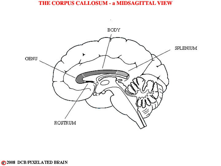
- - In summary, deep within the cerebral hemisphere lie a group of nuclei (groups of neuronal cell bodies packed together) called the basal ganglia. These nuclei include the caudate, the putamen, the globus pallidus, the amygdala and the claustrum. The migration of neural tissue, as the hemisphere has enlarged, has caused these nuclei (particularly the caudate and the associated lateral ventricle) to assume odd shapes. Threading between these nuclei are the pathways that connect the cerebral cortex with other regions of the brain. It is useful to be able to visualize the complex anatomy that results in order to interpret radiographic sections through the hemisphere.
- - Of course, you will never see these structures (in real life) as they have been depicted in the above views. Rather, you will see them in radiographic views which are sections through the brain in a variety of planes. To get an idea of what lies ahead look at Blumenfeld pages 106-109 for typical horizontal sections through the brain; then look at pages 110-111 for typical frontal (=coronal) sections. Then, leaf through your Atlas of Whole Brain Sections (handed out at the start of the course) to become familiar with this resource.
- - Finally, click on Figure 7-2 and on 45 in the green box below the figure to call up frontal slide 45. The view that comes up will be a labeled version of the slide, similar to the one in you atlas. Click on unlabeled in the blue box on the left. That will give you an unlabeled view. Toggle back and forth between the two and convince yourself that you can identify all the structures that we have discussed above. Then, do the same for slide 38. After that, click on Figure 7-3 and look at horizontal slides 4 and 8 in the same way.