PIXBRAIN HOME _ _ _ MOD 3 HOME _ _ _ previous_ _ FIGURE 3-4_ _ next _ _ _ I WANT TO
- - - The size of the receptive field of these neurons varies as a function of the part of the body's surface represented. One simple measure of this is the 2 point discrimination test. The patient is tested by touching his skin with a pair of dividers. The spread between the tips is gradually increased until the patient recognizes the stimulus as two points rather than one. This distance is a rough measure of the size of the receptive field of individual sensory neurons. The distance is smallest (3-4 mm) on the finger tips and the tongue. It is somewhat larger for the hands and peri-oral area, and larger still (by a factor of 10) for the arms.
- - - This means that the map of the body's surface projected on the brain is a distorted one with the fingers, hands, tongue and peri-oral surfaces over represented. At the other end of the scale, the back has the poorest representation.
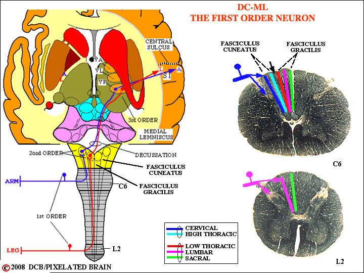
- - - The central processes of these first order neurons enter the spinal cord in the medial part of the dorsal root entry zone and bifurcate. One branch passes deeply into the dorsal horn and terminates in laminae III, IV, and V. For the most part, these fibers represent the afferent side of a variety of spinal reflexes. The other branch of the incoming fiber passes medially into the dorsal funiculus and ascends to terminate in either the nucleus gracilis (if it originated below the mid-thoracic level) or the nucleus cuneatus. These nuclei lie on the dorsal surface of the medulla, just above the cervical cord.
- - - The dorsal columns have a very precise somatotopic organization, but they show a segregation by function, as well. The axons of rapidly adapting neurons tend to lie more deeply and to terminate in the central and caudal parts of the relay nuclei. Slowly adapting neurons plus those associated with muscle spindles and joint receptors terminate within the rostral pole of the nucleus. This type of segregation is maintained all the way to the cortex