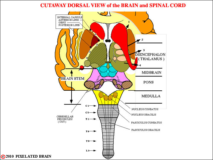
FIGURE 3-1
This view is similar to that of Figure 2-4, except that the numbers denoting slide levels have been removed and labels showing the regions of the brainstem (and the diencephalon) have been added. In addition, numbers and arrows have been added to help us describe the internal capsule. On the right, the numbered arrows show how fibers radiate laterally from the thalamus, passing "under" the caudate, and entering the internal capsule. On the left, a horizontal cut has been made through the thalamus, revealing the classic "V" shape of the internal capsule. The "1" on both sides shows the position of fibers traveling in the anterior limb of the capsule; The "2, 3, 4" show the position of fibers traveling in the posterior limb.
Note the position of the nuclei and fasciculi associated with the DC-ML pathway.