PIXBRAIN HOME _ _ _ MOD 3 HOME _ _ _ previous_ _ FIGURE 3-2_ _ next _ _ _ I WANT TO
- - - The first neuron activated by the stimulus is called the "primary" or "first order" neuron. Activation of this neuron specifies both the "where" and "what kind of" aspects of the stimulus.
- - - In the case of touch, each primary neuron has a circumscribed "receptive field": the size of this field varies in a predictable manner, as discussed below
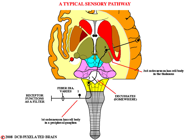
- - - Sensory information is projected to the cerebral cortex by synaptic relay through a series of neurons.
- - - Typically, there are said to be three neurons in the chain: the first order neuron, which always (with one minuscule exception) has its cell body in a ganglion outside the central nervous system; a second order neuron, having its cell body either in the brainstem or spinal cord; and a third order neuron with its cell body in the thalamus. The second and third order neurons are commonly found in "relay nuclei". While relay nuclei are often diagrammed as simple relay sites, a significant amount of processing of the neural message may take place within them. There may, for example, be interaction between the ascending sensory message and a descending input, originating in the cerebral cortex
- - - In most cases, sensory information is projected to the cortex by more than one pathway. Such "parallel pathways" often consist of a phylogenetically old one (or more) which convey a limited amount of information by a relatively slow route, and a newer pathway which conveys precise information by a rapid route. The anterolateral pathway (which we will discuss tomorrow) is an example of the former, and the dorsal column-medial lemniscus pathway is the premier example of the latter.
- - - Finally, many sensory pathways are organized to preserve the spatial relationships of the receptors in the periphery. We use terms like somatotopic, retinotopic and tonotopic to refer to this quality.