PIXBRAIN HOME _ _ MOD 4 HOME _ _ _ previous _ _ FIGURE 4-1__ _ next _ _ _ I WANT TO
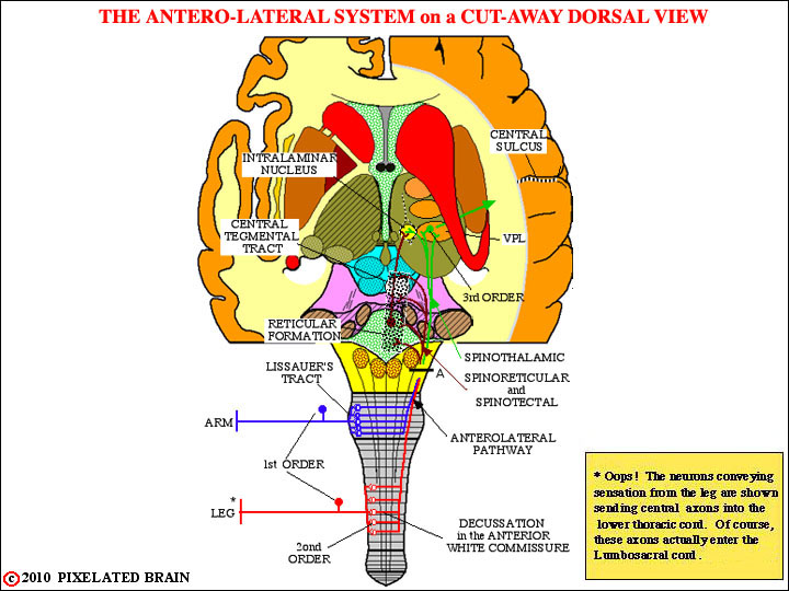
- - - The lower part of this figure, below line A, illustrates how the anterolateral pathway is formed at spinal cord levels.
- - - The axons of the first order neurons are relatively small in diameter A-delta or C fibers. They enter in the more lateral part of the dorsal root entry zone, and may travel up or down the cord for several segments in the dorsolateral fasciculus of Lissauer (Lissauer's Tract) before turn deeply to end on second order neurons in the dorsal horn. The manner of termination is complex, and somewhat different for each component of the system. The second order neurons give rise to axons that cross the midline in the anterior white commissure and ascend contralaterally in the anterolateral fasciculus. The pathway is somatotopically organized at spinal levels; the fibers that join the pathway as it ascends (the blue ones in the above figure) pile up on the deep (inner) side. While this somatotopic organization is clinically significant at spinal cord levels, it is not obvious once the pathway reaches the brainstem.
- - - The upper part of this figure, above line A, illustrates how the divisions of the anterolateral system ascend through the brainstem.
- - - The axons in this spinoreticular division of the anterolateral pathway do not ascend directly to the thalamus. Rather, they gradually "peel off", as the pathway passes through the brainstem, and terminate on neurons of the reticular formation. Cells of the reticular formation that have received a spinoreticular input relay this information via the central tegmental tract, to terminate in the intralaminar nuclei of the thalamus. The axons in this spinomesencephalic (spinotectal) division of the pathway pass somewhat more rostrally to end in the tectum of the midbrain. Fibers may also end in the periaqueductal gray region, just below the colliculus. The precise role of this pathway is uncertain. Some believe it is responsible for initiating eye movement in response to painful stimuli. Others suggest it represents the afferent or input side of a system which participates in the modulation of pain.
- - - The axons in this spinothalamic division of the anterolateral pathway ascend directly to the thalamus, terminating as shown in the figure above.