PIXBRAIN HOME _ _ MOD 8 HOME _ _ previous _ _ FIGURE 8-7_ _ next _ _ I WANT TO
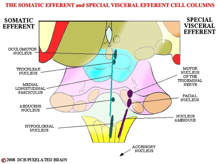
The oculomotor, trochlear and abducens nerves innervate the muscles that move the eyes. The medial longitudinal fasciculus serves to connect these nuclei and coordinate their activity. The hypoglossal nerve, of course, has nothing to do with eye movement - rather, it innervates the muscles of the tongue. These nuclei are all a part of the Somatic Efferent (SE) cell column and we take them up in Module 9
The four nucle of the Special Visceral Efferent (SVE) cell column are shown here in purple. The (spinal) Accessory nucleus lies in the upper 5 or 6 segments of the cord, indicated here by the arrow. While most of the cell columns, like the SE one above are at best, interrupted ones, the SVE column is really an almost continuous one. The motor neurons in these nuclei are typical "lower motor neurons", just like those in the spinal cord. What makes them "special" is that they innervate muscles of branchial arch origin. The muscles are striated, so many people are uneasy about calling them "special VISCERAL motor. To get around this they are often called Branchiomotor.
Just like spinal motor neurons, the SVE ones are driven by upper motor neurons in the lateral part of the precentral gyrus. This pathway departs from the corticospinal one at the midbrain level (Figure 5-5), so we can't call it part of the pyramidal tract (it doesn't go through the pyramids of the medulla). It is called the coriticobulbar pathway and you traced it in Module 5 (remember all those yellow dots). If you want a quick review, click on Slide 28, and then use the "caudal buttons" to follow it downward through the tegmentum. We discuss it later in the module.