PIXBRAIN HOME _ _ MOD 12 HOME _ _ previous _ _ FIGURE 12-1_ _ next _ _ I WANT TO
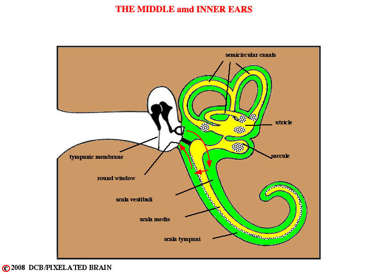
Variations in air pressure (i.e. sound) pass through the external acoustic meatus to reach the tympanic membrane. The meatus, a canal in the petrous part of the temporal bone, is shown (but not labeled) above. Movement of the tympanic membrane is transmitted by the bones of the middle ear to the foot plate of the stapes, positioned within the oval window of the inner ear . This in turn causes deflections of the basilar membrane, which travel from the base to the apex of this structure. The basilar membrane is "tuned" in such a way that low frequencies resonate near the apex and high frequencies resonate near the base. However, the mechanical tuning of the membrane is rather broad - particularly for low frequencies - and cannot, by itself, account for the way in which sound waves are so precisely translated into a neural code. Read the section on neural coding in Kingsley (Pg.. 408-412) for a discussion of this issue. When the basilar membrane moves, the hair cells that lie on it are alternately hyperpolarized and depolarized; depolarization initiates action potentials in the peripheral terminals of the first order neurons of the auditory pathway. These neurons have their cell bodies in the spiral ganglion, buries within the modiolus, and send their central processes through the internal acoustic meatus to enter the cranial cavity.