PIXBRAIN HOME _ _ HOME MOD 1 or 2 _ _ previous _ _ Lect-Dev - Fig 6 _ _ next _ _ I WANT TO
- - - - - Like the brain, the spinal cord initially forms by a rolling up of the neural plate to form the neural tube. The neuroepithelial cells of the tube's walls will generate all of the neurons and glia of the spinal cord.
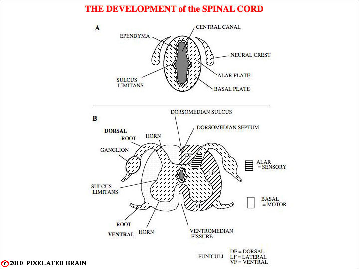
Early in development there is a slight indentation in the wall of the neural tube, the sulcus limitans, which divides the two major zones of proliferation, the dorsal alar and ventral basal plates. Neurons derived from the alar plate are primarily ascending projection neurons and interneurons involved with sensory function in the dorsal horn. Neurons in the basal plate differentiate into the motor neurons and interneurons of the ventral horn of the spinal cord.
- - - - - The mature spinal cord resembles a butterfly, with the central gray matter containing many neuronal and glial cell bodies, and the peripheral white matter containing the myelinated fiber tracts of ascending and descending axons. As described above, neurons in the gray matter of the dorsal horn are mainly involved with processing sensory information which enters the spinal cord through the dorsal root from the dorsal root ganglion neurons. Neurons in the ventral horn provide mainly motor output. Their axons leave the spinal cord through the ventral root.
- - - - - The peripheral layer of axons which forms the white matter is grossly divided into three bundles by the dorsal and ventral roots. These bundles are referred to as the dorsal, lateral and ventral funiculi.