DEV-8
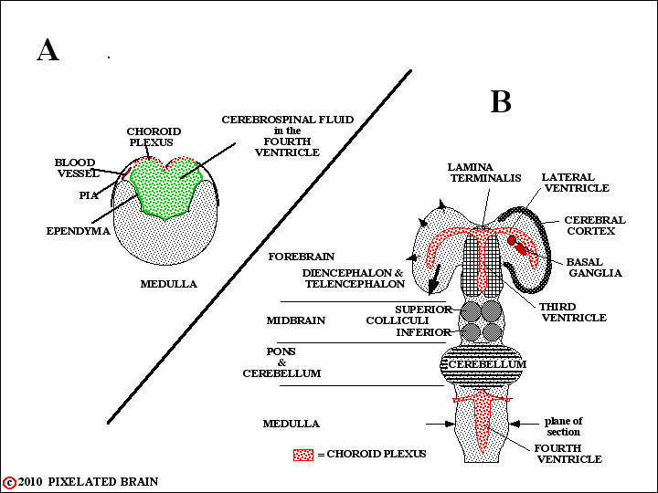
In some regions the edges of the neural plate fail to meet. In these instances , the ependymal cell layer, lining the cavity, and the layer of pial cells, covering the surface of the tube, meet to form a membrane which completes closure of the cavity. Blood vessels extend into this membrane creating highly vascular tissue called choroid plexus. The two regions where this normally occurs are shown in this highly schematic figure. In A, which is simply a restatement of Figure 7, we see the situation in the medulla. In B, which is a restatement of Figure 8, we see that the edges of the neural tube also fail to meet at the level of the diencephalon. Thus both the third and fourth ventricles are roofed by choroid plexus. Furthermore the choroid plexus of the third ventricle is carried out into the lateral ventricles within the hemisphere. Choroid plexus tissue is the main source of CSF, and since the greatest mass of this tissue is found lining the two lateral ventricles this is where most CSF is produced.