PIXBRAIN HOME _ _ MOD 1 HOME _ _ previous _ _ FIGURE 1-8 _ _ next _ _ I WANT TO
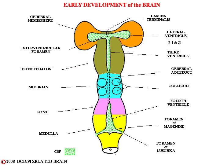
- - As development of the brain continues, regional variations in the tube become apparent. The most rostral region (the diencephalon) and the most caudal (the pons and medulla) contain ventricles roofed by choroid plexus. The intermediate part (the mid brain) is a true tube having a central canal called the cerebral aqueduct. The ventricular system is closed off rostrally by the lamina terminalis. With time, the wall of the diencephalon adjacent to the lamina terminalis evaginates laterally on either side, carrying the central cavity along with it, to form the cerebral hemisphere. Choroid plexus is also carried out into these new ventricles where it seals off a rather extensive defect in the ventricular wall. The ventricles within the hemisphere are named the lateral ventricles. They are considered the first two ventricles of the brain, with the result that the cavity of the diencephalon is called the third ventricle and the one in the caudal brainstem is named the fourth ventricle. The constricted channel through which the lateral ventricle communicates with the third one is the inter ventricular foramen (of Monro). Choroid plexus tissue is the main source of CSF, and since the greatest mass of this tissue is found lining the two lateral ventricles this is where most CSF is produced.