PIXBRAIN HOME _ _ MOD 2 HOME _ _ previous _ _ FIGURE 2-2 _ _ next _ _ I WANT TO
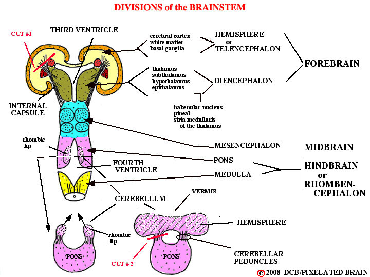
- - The various names applied to each part of the brain are given in this view.
- - You are already familiar with the way in which the cerebral hemisphere develops by evaginating outward from the side wall of the diencephalon. The cerebellum, in contrast, develops as a proliferation of the cells in the edge of the neural fold (the so-called rhombic lip, because it forms a margin of the rhomboid - shaped fourth ventricle) at the pontine level. The cell masses on either side join in the midline to become the cerebellum. Unlike the cerebral hemisphere, the cerebellum has a midline component (the vermis) as well as two lateral extensions, the cerebellar hemispheres. Both the cerebral hemisphere and the cerebellum are "anchored" to the brainstem by massive bundles of fibers (axons) which are, in fact, the input and output pathways of these structures. In the case of the cerebral hemisphere, this fiber bundle is the internal capsule, which you have already studied. The cerebellum has 3 separate fiber bundles of this sort, known as the cerebellar peduncles.
- - In any attempt to look at the dorsal and lateral surfaces of the brainstem, the first problem to be confronted is that this region is hidden below the cerebral hemispheres and cerebellum. You will recall that the expanding cerebral hemisphere extends caudally to completely cover the diencephalon and midbrain . How can we expose the brainstem to view? The obvious answer is to dissect away the tissue of the cerebral hemisphere (and of the cerebellar hemisphere, as well) which gets in the way. The most "pure" approach , at least in conceptual terms, would be to cut the relevant fiber bundles and then just lift off the hemispheres (cerebral and cerebellar). A careful look at this figure shows where to make the cuts. After making cut #1 (and this is pretty schematic) you would be able to lift off the cerebral hemisphere, leaving behind the basal ganglia, the diencephalon and the brainstem. After making cut #2 you would be able to lift off the cerebellum. The result would be to give you a good exposure of the dorsal surface of the brainstem (plus the basal ganglia and diencephalon). In fact, this is exactly what we propose to do.