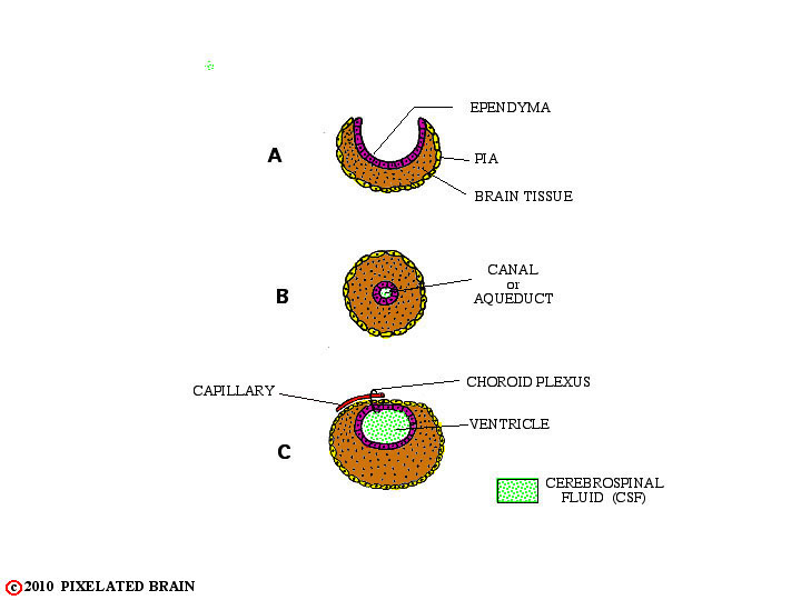
FIGURE 1-7
In two regions, however, the edges of the neural plate fail to meet, as shown A. In these instances , the ependymal cell layer, lining the cavity, and the layer of pial cells, covering the surface of the tube, meet to form a membrane which completes closure of the cavity. Blood vessels extend into this membrane creating highly vascular tissue called choroid plexus (see B and C, above). The two regions where this occurs are shown in the highly schematic Figure 8.