PIXBRAIN HOME _ _ MOD 1 HOME _ _ previous _ _ FIGURE 1-19 _ _ next _ _ I WANT TO
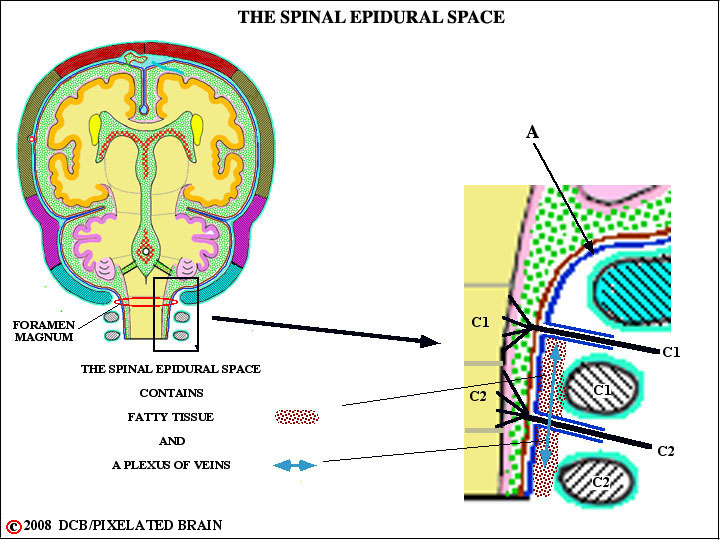
- - At the Foramen Magnum the periostial and meningeal layers of the dura part ways (arrow A). The periostial layer continues on to the external surface of the skull, while the meningeal layer forms a tube, hanging rather freely within the vertebral canal, enclosing the spinal cord. Since the cranial dura is tightly adherent to the skull, there is - normally - no epidural space in the cranial cavity. In contrast, there is a true spinal epidural space and, as shown here, it is filled with fatty tissue and a plexus of veins. The venous plexus, known as Batson's plexus, is important because it extends the length of the vertebral canal and the veins have no valves, allowing blood flow in either direction. This forms an important route by which prostatic cancer cell may easily spread to vertebrae.
- - The dura forms a pressure tight seal around everything that passes through it - blood vessels and nerves (shown here) - with the result that the pressure within the cranial cavity and spinal dural sleeve differs from atmospheric pressure. For a description of how this pressure is measured, see Blumenfeld, pgs. 161-2/ Fitzgerald, pgs. 49-50.