PIXBRAIN HOME _ _ MOD 1 HOME _ _ previous _ _ FIGURE 1-15 _ _ next _ _ I WANT TO
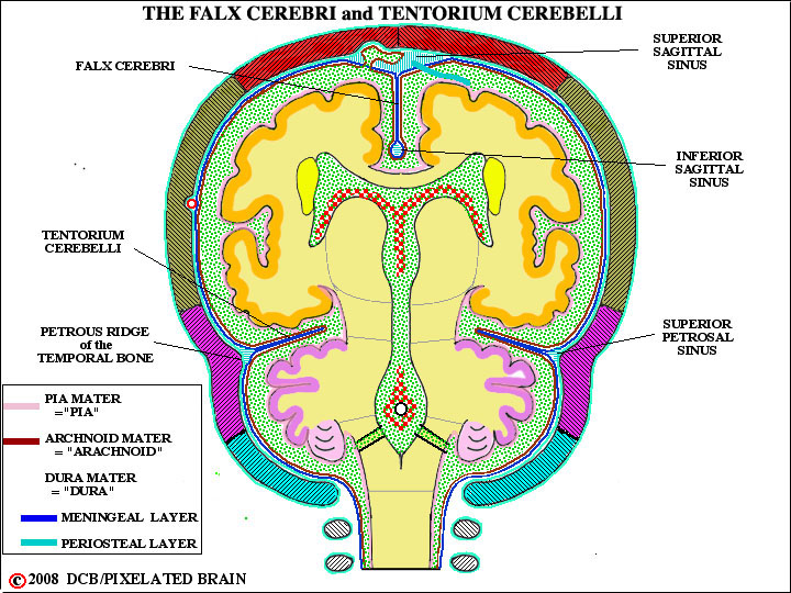
- - In three regions the meningeal layer of the dura breaks away from the dural one to form tough, unyielding folds or partitions which partially subdivide the cranial cavity. Two of these - the falx cerebri and the tentorium cerebelli - are shown here. The falx cerebri, and its posterior extension, the falx cerebelli, is also shown in Figure 16. The third dural fold is the diaphragm sella,which partially roofs the hypophyseal fossa, leaving a small opening for the stalk of the pituitary (see Figure 1-21). These partitions are important because when expanding lesions in the cranial cavity - blood or a tumor - displace brain tissue, it is often obliged to herniate around one of them. For a discussion of these herniation syndromes see Blumenfeld, pg. 140 / Fitzgerald, pg. 77 / Haines pg. 111.
- - When the dural layers separate, as described above, they often enclose a network of venous sinuses. This is shown most clearly in this view, where the superior sagittal sinus. the inferior sagittal sinus and the superior petrosal sinus are labeled. Another example, in which the meningeal dura encloses a sinus, is the straight sinus (Figure 16). In this, the sinus is created in the region where the falx cerebri joins the ridge line of the tentorium cerebelli. See Fitzgerald, pgs. 44-45 or Haines, pgs.110-112 for more diagrams and details.