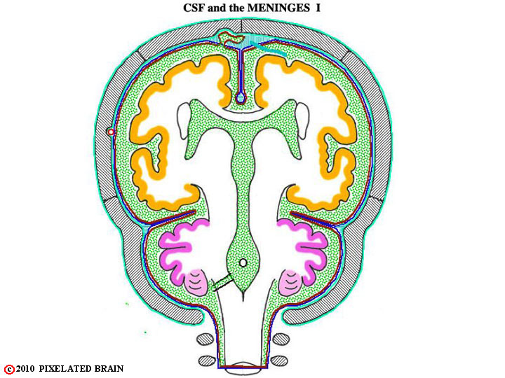
FIGURE 1-9a
This is a schematic section through the brain that will be useful in describing the flow of CSF and the meninges. Use it (as well as the other figures of this group), to confirm the following:
1) The dura may be thought of as a two layered structure, with an outer periostial layer (which serves as the periosteum of the skull) and an inner meningeal layer. The periostial layer is tightly adherent to the overlying bone, and in most regions the two layers of the dura are fused.
2) The two layers split to enclose the middle meningeal artery. The artery is positioned between the two layers in such a way that it creates a ridge on the outer (periostial) surface of the dura, but leaves the inner (meningeal) surface quite smooth (Figures. 1-7 & 1-8). One of the results of this arrangement is that the artery "erodes" a valley on the inner surface of the skull. Because skull fractures which cross this valley may well tear the artery and lead to intracranial bleeding, the precise course of the artery is of clinical significance.3) The two layers also split to enclose the venous sinuses of the cranial cavity. Three of these - the superior sagittal sinus, the inferior sagittal sinus and the straight sinus - are shown in Figure 1-9. Others you should be aware of include the transverse sinus, the sigmoid sinus and the superior and inferior petrosal sinuses.
4) The meningeal layer of the dura evaginates, or folds in upon itself, to create several partial partitions within the cranial cavity. These include the falx cerebri (Fig, 1-9), the falx cerebelli (Figure 1-10) and the tentorium cerebelli.
5) At the foramen magnum the two layers of dura separate (Figure 1-10); the endosteal layer passes around to the external surface of the skull while the meningeal layer descends within the spinal column to form a covering of the spinal cord.
6) The dura is sensitive to pain. Trigeminal fibers supply the dura of the anterior and middle cranial fossae, as well as the tentorium. The vagus nerve and cranial nerves 2 and 3 supply the dura of the posterior cranial fossa.
When the dura is lifted off the brain, the next meningeal layer, the arachnoid, is exposed. The two layers separate easily except in the region of the superior sagittal sinus. Here, two structures impede the separation: 1) "bridging veins", draining the surface of the hemisphere, pass through the arachnoid to empty into the sinus and 2) small evaginations of arachnoid, termed arachnoid granulations, push their way into the interior of the sinus. Arachnoid granulations represent the sites where cerebrospinal fluid, secreted into the ventricles of the brain, is returned to the venous system. We will return to the topic of cerebrospinal fluid later.