PIXBRAIN HOME _ _ MOD 8 HOME _ _ previous _ _ FIGURE 8-3_ _ next _ _ I WANT TO
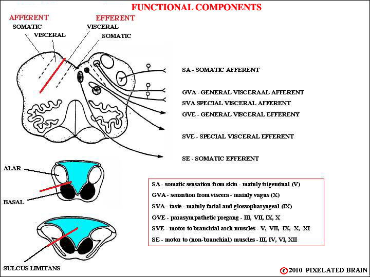
You will remember (we hope) that when the neural plate rolled up to form a tube the sulcas limitans served to form a marker, seperating the dorsal "alar plate" from the ventral "basal plate" as seen in emb_fig 06 . There are, however, regions where the lips of the neural plate don't meet; in these regions the roof of the central cavity is sealed by choroid plexus tissue, and the alar plate tends to lie lateral to the basal plate, rather than dorsal to it - as shown above and in emb_fig 08 . The picture is further complicated by the appearance of four new functional groupings, serving functions unique to the head. These are thought of as "visceral" because the are associated with the rostral end of the gut or alimentary canal. As the diagrams show, the sensory nuclei lie dorsolateral to the sulcus limitans and the motor nuclei lie ventromedial to it.
The sulcus limitans is only obvious in the caudal medulla, but a suggestion of it is also present in the diencephalon where a slight "valley" separates the dorsal thalamus (which might be thought of as a sensory structure) from the ventrally placed hypothalamus (which might be thought of as a motor structure).
Blumenfeld illustrates this topic it in his Figure 12.4.