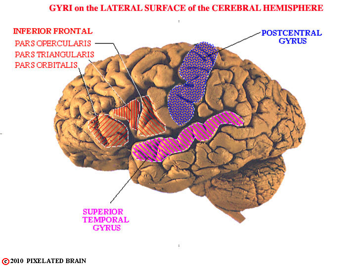
FIGURE 1-33
Most textbook figures imply that the pattern of gyri and sulci on the surface of the hemisphere is quite similar from one brain to the next. In reality, there is a great deal of variability between specimens. In the brain we looked at originally, for example, the central, post central and superior temporal sulci are rather clear, with the result that the post central gyrus and the superior temporal gyrus are easily identified, as shown here. In other brains, even the central sulcus (the most obvious of the three) can be difficult to identify.