FIGURE 2-9
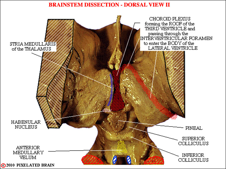
This view shows the course of the choroid plexus and identifies other structures. Switch back and forth between these copies and be certain you can identify the following:
1) the septum pellucidum: This thin membrane runs between the corpus callosum and the fornix and separates the anterior horns of the two ventricles from each other
2) the caudate nucleus
3) the stria medullaris of the thalamus. This is a longitudinal bundle of axons which also marks the point of attachment of the choroid plexus that normally roofs the third ventricle (but is removed in this dissection)
4) the habenular nucleus.
5) the pineal
6) the anterior medullary velum. This membrane forms the roof of the anterior part of the fourth ventricle.
7) the superior and inferior colliculi