_ _PIXBRAIN HOME _ _ MOD 1 HOME _ _ previous _ _ FIGURE 1-17 _ _ next _ _ I WANT TO
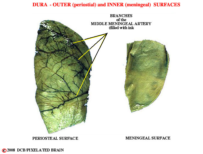
- - The dura, shown in Figure 1-7, was subsequently removed from the surface of the brain and cut into these two pieces. Then india ink was injected into the middle meningeal artery. Note how the branches of the vessel stand out on the outer ( = periosteal ) surface. If you rubbed your finger over it, you would easily feel these ridges. In contrast, the inner ( = meningeal ) surface is quite smooth. When the brain moves slightly within the skull (when you bump your head against a wall, for example) the brain's surface moves, relative to the inner surface of the dura, and it makes sense for the latter to be smooth. The result of this asymmetrical split is that the artery erodes a "valley" in the inner surface of the skull., as shown in Figure 18. If the skull fracture and the fracture line crosses this valley, then there is a good chance the middle meningeal artery will be torn and that arterial blood will leak forming a hematoma (pool of blood) between the dura and the skull.