A SURVEY of the PIXELATED BRAINSTEM SLIDE SERIES
There are 38 slides (#1-#38) in this series; because of the bend in the axis of the brain at the midbrain level, the sections pass somewhat obliquely through the brainstem, but are close to "true" frontal sections through the diencephalon. We have organized them into 8 regions in this presentation. On the left are groups of 4-6 adjacent sections. To the right of each group is a list of 10 features common to most of the sections in the group. While the sections are unlabeled, each one is a "hot spot". Clicking on it carries you to 2 labeled versions of the same slide.
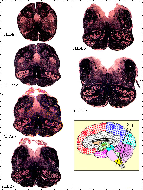 Figure 1
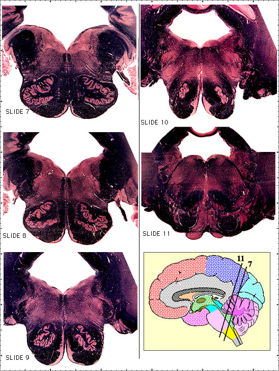
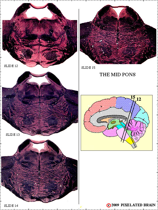 Figure 3
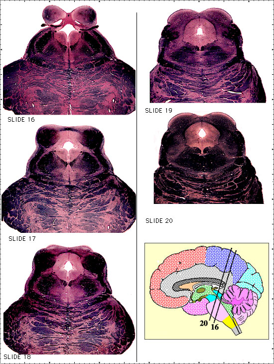 Figure 4
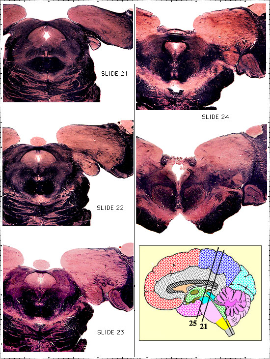 Figure 5
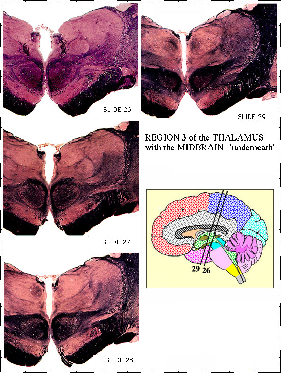 Figure 6
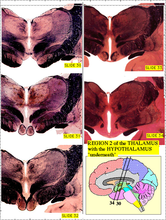 Figure 7
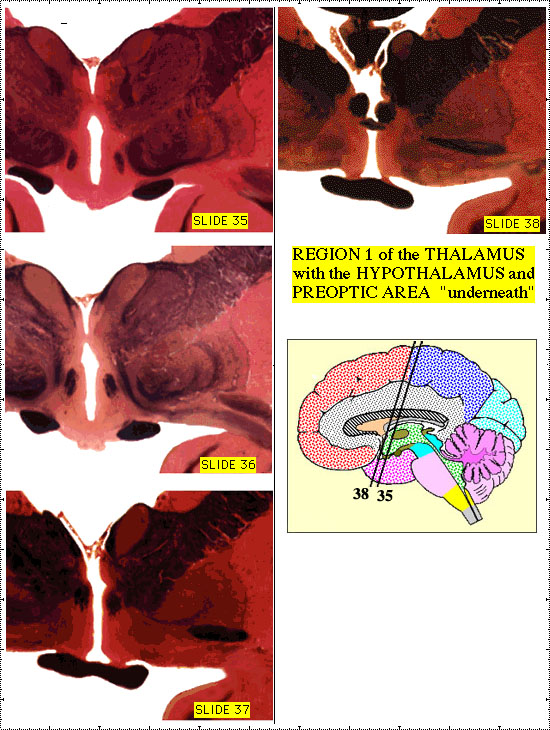 Figure 8
THE MEDULLA
1) the central canal of the spinal cord opens into the 4th ventricle
2) fibers originating in the nuclei gracilis and cuneatus decussate as internal arcuate fibers to form the medial lemniscus
3) the pyramid (containing descending corticospinal fibers) and inferior olive are prominent landmarks, present in most sections
4) nuclei associated with the hypoglossal and vagus nerves are found here, and rootlets of these nerves exit on either side of the inferior olive
5) fibers originating in the inferior olive decussate and are joined by those of the dorsal spinocerebellar tract to form the inferior cerebellar peduncle
6) the cerebellum lies dorsal to the medulla // the space between these two structures is occupied by the 4th ventricle // the deep nuclei of the cerebellum are present at this level
7) the spinal tract of the trigeminal nerve descends through this region to reach the cervical cord // the nucleus of the tract always lies medial to it
8) the foramen of Luschka marks the anterior border of the region // rostral to this the inferior cerebellar peduncle attaches the brain stem to the cerebellum
9) the medullary reticular formation is present throughout this region, but is shown only in slide #5
10) the vestibular nerve enters more rostrally but nuclei associated with it (vestibular nuclei) descend in this region
THE MEDULLA/PONS JUNCTION
1) in this transitional zone, the ventral part of most sections "looks like" the medulla (pyramid and inferior olive present) and the dorsal part "looks like" the pons (brain stem attached to cerebellum)
2) this is the level of the cerebello-pontine angle // the facial, vestibulocochlear and glosso-pharyngeal nerves exit laterally (in the "angle") and the flocculus forms the lateral border of the angle
3) cerebellar peduncles are prominent // the inferior and middle peduncles enter the cerebellum, and the superior one begins its exit // all 3 are present in some sections
4) nuclei associated with the facial and vestibulocochlear nerves are found at this level
5) the spinal tract of the trigeminal nerve descends through this region to reach the cervical cord // the nucleus of the tract always lies medial to it
6) the medial lemniscus seems trapped in a vertical position between the two inferior olives, but begins to slide laterally once it gets rostral to these nuclei
7) the 4th ventrical is largest at this level // it is roofed by the cerebellum and, at rostral levels, by the anterior medullary velum
8) the fibers originating in the facial nucleus take an unusual route (the internal genu) as they loop around the abducens nucleus to exit
9) the pontine reticular formation is present at all levels, but is shown only for slide 10
10) the occipital lobe of the cerebral hemisphere seems to lie on the surface of the cerebellum, but is actually separated from it by the tentorium cerebelli
THE MID PONS
1) this zone is "owned" by the trigeminal nerve // the fibers of the nerve pass medially through the middle cerebellar peduncle to reach the chief sensory and motor nuclei in slide 12
2) the fibers of the spinal tract turn caudally to descend to the cervical cord // the fibers of the mesencephalic tract turn rostrally to reach the midbrain
3) the 4th ventricle diminishes in size at the rostral end of this zone and becomes the cerebral aqueduct
4) the medial lemniscus is now free of the inferior olive and assumes a horizontal position as it moves laterally
5) the fibers of the ascending auditory pathway cross the midline just ventral to the medial lemniscus forming a region called the trapezoid body
6) the superior cerebellar peduncle gradually wraps around the 4th ventricle to enter the tegmentum of the brain stem at the rostral border of the region // the cerebellum diminishes in size // the tentorium still lies
between it and the occipital lobe of the cerebral hemisphere
7) the medial longitudinal fasciculus passes rostrally through this level to reach the oculomotor and trochlear nuclei in the midbrain
8) neurons of the reticular formation contribute axons to the central tegmental tract, which ascends through the tegmentum
9) within the massive basalar pons, pontine nuclei send axons across the midline to form the middle cerebellar peduncle and enter the cerebellum
10) the corticospinal tract is now buried within the basilar pons
THE MIDBRAIN -PONS JUNCTION
1) the pontine tementum lies in the middle of each section // dorsal to it is the tectum of the midbrain, and ventral to it is the basilar pons
2) the caudal 2 slides pass through the round, inferior colliculus and the rostral 2 through the more oval superior colliculus // the middle slide of the group shows parts of both colliculi
3) the cerebellum disappears and the most rostral slides cut tangentially through the pulvinar of the thalamus
4) the superior cerebellar peduncle moves medially through the tegmentum and finally decussates at the rostral border of the region
5) we can no longer ignore the cerebral hemisphere // many structures assume a "C" shape as they are "pulled out" into the temporal lobe and 3 of them are cut tangentially or twice in these sections // they are the
lateral ventricle, the fornix and the caudate nucleus
6) the medial "edge" of the cortex of the temporal lobe is folded upon itself in a complex manner to form the hippocampus
7) the corpus callosum appears, connecting the two cerebral hemispheres // at this level, some of the more ventral fibers form a bundle that connects the two hippocampi (the hippocampal commissure)
8) the auditory pathway can be seen approaching the inferior colliculus as the lateral lemniscus // more rostrally, it departs from the colliculus as the brachium of the inferior colliculus
9) the tentorium is still present, but now has a "free edge" // it is through the limited opening medial to this edge that the brainstem extends rostrally // the opening framed by the tentorium is termed the "tentorial notch"
10) the cerebral aqueduct, surrounded by the central gray, is one of the "hallmarks" of the midbrain // the colliculi are another
REGION 4 of the THALAMUS with the
MIDBRAIN (mainly) "underneath"
1) the dorsal part of each section passes through the thalamus , one of the subdivisions of the diencephalon
2) the thalamus is an "egg shaped" area, packed full of nuclei // we divide it into 4 regions and these sections pass through region #4 // three nuclei are present in this region - the pulvinar, the lateral geniculate nucleus and the medial geniculate nucleus
3) the ventral part of these slides gradually (at more rostral levels) assumes the characteristic appearance of the ventral midbrain // the defining structures are the red nucleus, the substantia nigra and the cerebral peduncle
4) the posterior commissure is one landmark defining the border between midbrain and diencephalon // just rostral to it the cerebral aqueduct opens into the 3rd ventricle // the habenular nuclei (of the diencephalon) flank this opening
5) the superior cerebellar peduncle completes its decussation, passing through and around the red nucleus as it continues to ascend toward the thalamus
6) the medial lemniscus moves laterally, also preparing to enter the thalamus
7) fibers of the corticospinal tract emerge from the basilar pons and lie within the cerebral peduncle
8) the oculomotor nucleus (a midbrain structure) is present but, because of the oblique plane of the slides, the exiting fibers of the nerve are not yet apparent
9) in the temporal lobe, the hippocampus assumes its characteristic "profile", and pushes into the inferior horn of the lateral ventricle
10) the 3 "C" shaped structures described previously are joined by a 4th - the stria terminalis // all of them (caudate, fornix, stria terminalis and lateral ventricle) are cut twice
REGION 3 of the THALAMUS with the
MIDBRAIN "underneath"
1) the dorsal part of each section passes through region #3 of the thalamus // the nuclei of the region are shown in the box that is a part of each slide view
2) the ventral part of each section has the characteristic appearance of the ventral midbrain // the defining structures are the red nucleus, the substantia nigra and the cerebral peduncle
3) the ascending fibers of the medial lemniscus enter the ventralis posterolateralis nucleus at the caudal border of this region // within this nucleus somatic sensory information is relayed on to the cerebral cortex
4) many of the fibers which ascended within the superior cerebellar peduncle now continue on to the thalamus in the cerebellothalamic pathway // to reach their relay nucleus, they begin to move laterally
5) the third ventricle is present // at this level, it completely divides the diencephalon into two halves, and it is roofed by choroid plexus
6) the oculomotor nerve exits from the midbrain, passing through the interpeduncular fossa
7) the lateral geniculate nucleus (a region #4 structure) is gone, but the input pathway to the nucleus - the optic tract - lies on the surface of the cerebral peduncle, laterally
8) throughout the region, fibers of the cerebral peduncle begin to pass dorsolaterally to become part of the internal capsule // this structure forms a "capsule" around the diencephalon // it provides a route by which pathways descending from the cerebral cortex to the brain stem and spinal cord can "bypass" the thalamus
9) structures in the temporal lobe are more or less unchanged, but the hippocampus becomes increasingly convoluted // the term hippocampal formation is used to include the hippocampus, the dentate gyrus and the subiculum
10) above the diencephalon lies the corpus callosum, with the fornix attached to its "under" surface // while the lateral and third ventricles may seem connected at this level, choroid plexus forms two partitions which separate them
REGION 2 of the THALAMUS with the
HYPOTHALAMUS "underneath"
1) because the axis of the brain bends sharply at the midbrain level, these slides are close to true cross-sections, rather than oblique cuts // as a result, they pass through a single region - the diencephalon
2) dorsally, the thalamic nuclei of region #2 are present
3) ventrally, the presence of the mammillary body in most slides establishes that they pass through the posterior region of the hypothalamus
4) the two halves of the diencephalon make limited contact (in some but not all brains) forming a structure called the massa intermedia
5) the structures (including the inferior horn of the lateral ventricle) which have been running forward in the temporal lobe now reach their anterior limits // just rostral (anterior) to this a large nucleus appears // it is the amygdala
6) lateral to the internal capsule two basal ganglia nuclei - the putamen and the globus pallidus - are evident // the globus pallidus sends fibers through (the fasciculus lenticularis) and below (the ansa lenticularis) the internal capsule to reach the thalamus
7) a thin nuclear area, the claustrum, separates the putamen from the cortex of the insula
8) the optic tract moves medially over the surface of the brain
9) between the thalamus and the hypothalamus lies the subthalamus, represented by a single nucleus - the subthalamic nucleus
10) cerebellothalamic fibers enter their thalamic relay nucleus - ventralis lateralis
REGION 1 of the THALAMUS with the
HYPOTHALAMUS and PREOPTIC AREA
"underneath"
1) these slides pass through region #1 of the thalamus // underneath it is either the hypothalamus or its rostral extension, the preoptic area
2) the putamen and globus pallidus now reach their maximum size // a third basal ganglion nucleus, the caudate, also is becoming larger as it pushes medially into the lateral ventricle
3) the ansa lenticularis completes its journey, passing underneath the internal capsule to reach the thalamus
4) the ventral amygdalofugal pathway takes a course similar to that of the ansa, connecting the amygdala with the diencephalon
5) the fibers of the optic tract reach the midline // half of them cross in the optic chiasm
6) while the anterior commissure crosses the midline just rostral to this region, its continuation out into the temporal lobe is evident here
7) the rostral border of the thalamus roughly marks the end of the original neural tube // it is here that lateral evaginations of the tube form the two cerebral hemispheres // thus it is in this region that the third ventricle communicates with the lateral ventricles, through the interventricular foramina
8) the fornix is cut twice as it arches downward and passes through the hypothalamus
9) the amygdala is still present, but begins to diminish in size
10) the choroid plexus of the third ventricle becomes continuous with that of the lateral ventricle in the region of the interventricular foramen
SITE MAP - - PIXBRAIN HOME - - I WANT TO