MODULE 4 - SECTION 3 - THE ANTEROLATERAL PATHWAY

29
|
MODULE 4 - SECTION 3 - THE ANTEROLATERAL PATHWAY
|
||
 |
 |
|
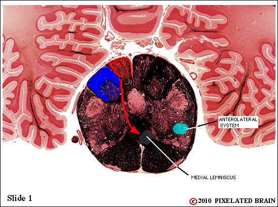
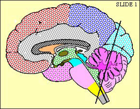
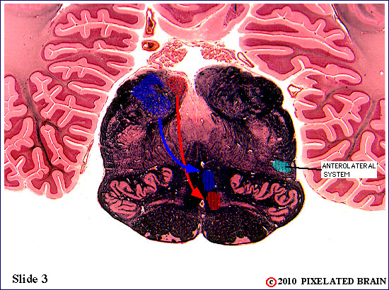
| The anterolateral pathway forms a reasonably compact bundle of fibers that ascend through the lateral part of the medula. Fibers of the spinoreticular division begin to "peel off" at this level and terminate on cells of the medullary reticular formation. While the spinothalamic division may retain some somatotopic organization, it is not precise enough to be of clinical significance. | ||
 |
 |
|
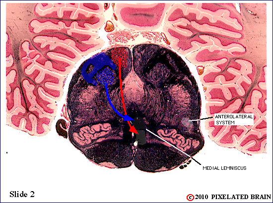
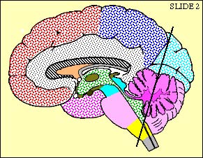
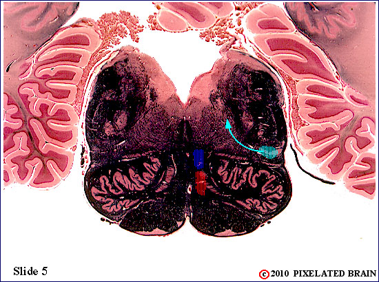
| The anterolateral pathway forms a reasonably compact bundle of fibers that ascend through the lateral part of the medula. Fibers of the spinoreticular division continue to "peel off" at this level and terminate on cells of the medullary reticular formation. While the spinothalamic division may retain some somatotopic organization,it is not precise enough to be of clinical significance. | ||
 |
 |
|
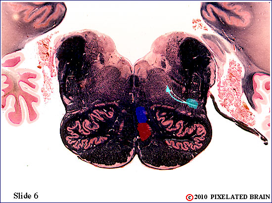
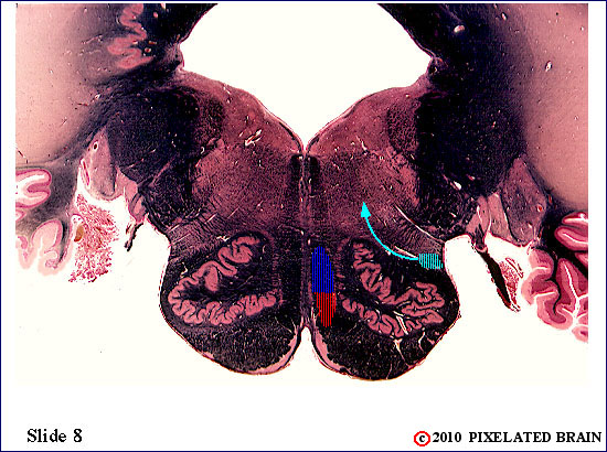
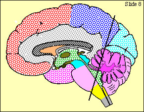
| The anterolateral pathway forms a reasonably compact bundle of fibers that ascend through the lateral part of the medula. Fibers of the spinoreticular division continue to "peel off" at this level and terminate on cells of the medullary reticular formation. While the spinothalamic division may retain some somatotopic organization, it is not precise enough to be of clinical significance. | ||
 |
 |
|
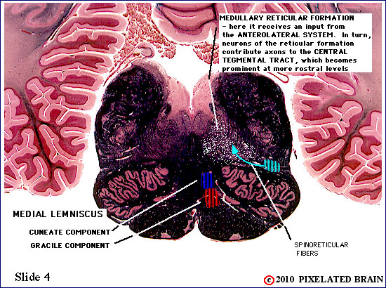
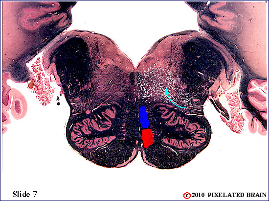
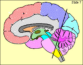
| The anterolateral pathway forms a reasonably compact bundle of fibers that ascend through the lateral part of the medulla. Fibers of the spinoreticular division continue to "peel off" at this level and terminate on cells of the medullary reticular formation. Cells of the reticular formation are scattered diffusely throughout the region indicated by the white stippling. These cells, in turn, give rise to axons which ascend to the thalamus within the central tegmental tract. While the spinothalamic division may retain some somatotopic organization, it is not precise enough to be of clinical significance. | ||
 |
 |
|
 |
 |
|
 |
 |
|
 |
 |
|
 |
 |
|
 |
 |
|
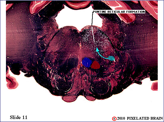
| The anterolateral pathway forms a reasonably compact bundle of fibers that ascend through the lateral part of the brainstem. Fibers of the spinoreticular division cotinue to "peel off" at this level and terminate on cells of the pontine reticular formation. Cells of the reticular formation are scattered diffusely throughout the region indicated by the white stippling. These cells, in turn, give rise to axons which ascend to the thalamus within the central tegmental tract. While the spinothalamic division may retain some somatotopic organization, it is not precise enough to be of clinical significance. | ||
 |
 |
|
| The anterolateral pathway forms a reasonably compact bundle of fibers that ascend through the lateral part of thepons. Fibers of the spinoreticular division still "peel off" at this level and terminate on cells of the pontine reticular formation. Cells of the reticular formation are scattered diffusely throughout the region indicated by the white stippling. These cells, in turn, give rise to axons which ascend to the thalamus within the central tegmental tract. While the spinothalamic division may retain some somatotopic organization, it is not precise enough to be of clinical significance. | ||
 |
 |
|
| The anterolateral pathway forms a reasonably compact bundle of fibers that ascend through the lateral part of the pons. Fibers of the spinoreticular division "peel off" at this level and terminate on cells of the pontine reticular formation. These cells, in turn, give rise to axons which ascend to the thalamus within the central tegmental tract. While the spinothalamic division may retain some somatotopic organization, it is not precise enough to be of clinical significance. | ||
 |
 |
|
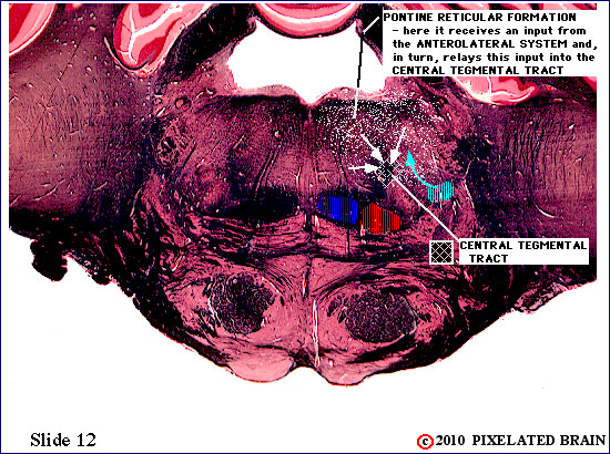
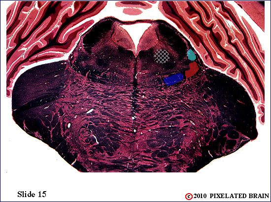
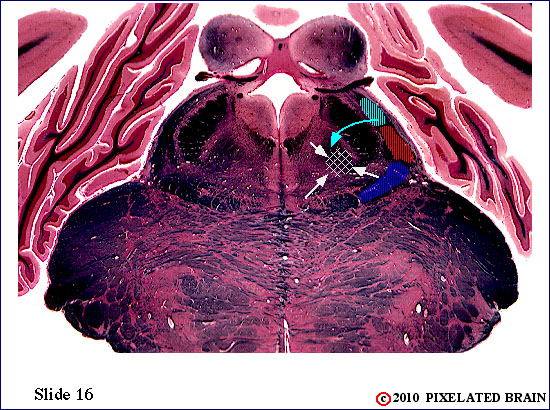
| The anterolateral pathway forms a reasonably compact bundle of fibers that ascend through the lateral part of the pons. Fibers of the spinoreticular division "peel off" at this level and terminate on cells of the pontine reticular formation. These cells, in turn, give rise to axons which ascend to the thalamus within the central tegmental tract (marked here by cross-hatching). While the spinothalamic division may retain some somatotopic organization, it is not precise enough to be of clinical significance. | ||
 |
 |
|
| The anterolateral pathway forms a reasonably compact bundle of fibers that ascend through the lateral part of thepons. Fibers of the spinoreticular division "peel off" at this level and terminate on cells of the pontine reticular formation. These cells, in turn, give rise to axons which ascend to the thalamus within the central tegmental tract (marked here by cross-hatching). While the spinothalamic division may retain some somatotopic organization, it is not precise enough to be of clinical significance. | ||
 |
 |
|
 |
 |
|
| The anterolateral pathway forms a reasonably compact bundle of fibers that ascend through the lateral part of the brainstem. Fibers of the spinoreticular division continue to "peel off" at this level and terminate on cells of the pontine reticular formation. These cells, in turn, give rise to axons which ascend to the thalamus within the central tegmental tract (marked here by cross-hatching). While the spinothalamic division may retain some somatotopic organization, it is not precise enough to be of clinical significance. | ||
 |
 |
|
 |
 |
|
 |
 |
|
 |
 |
|
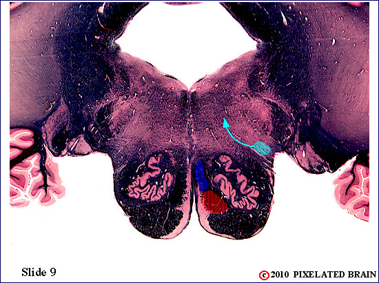
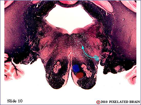
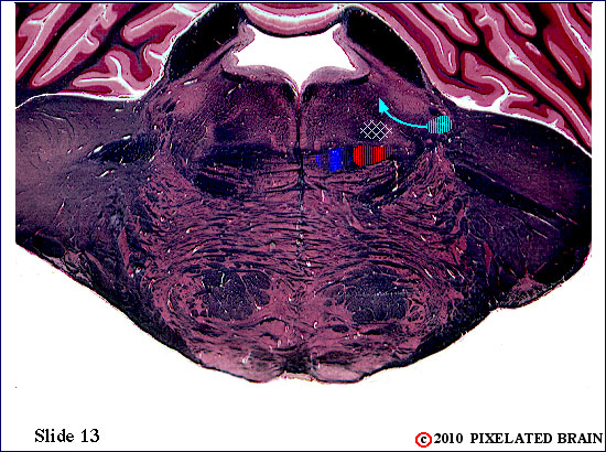
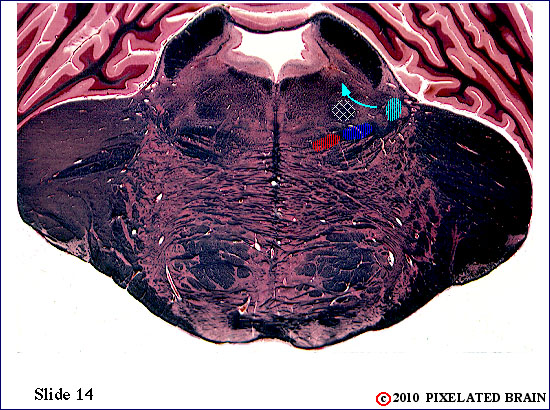
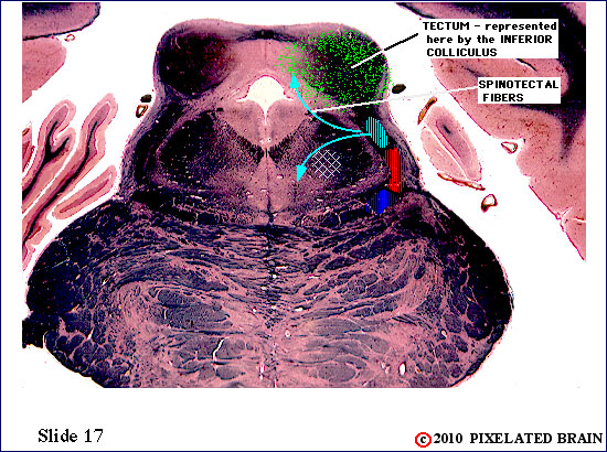
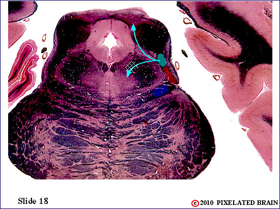
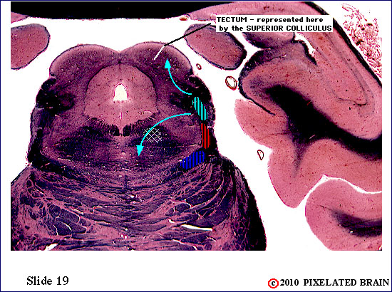
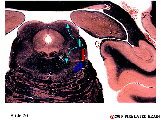
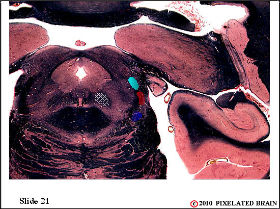
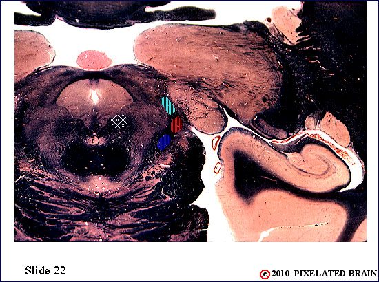
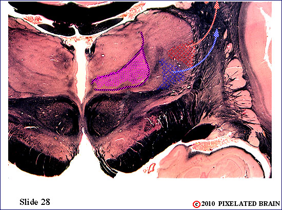
| The anterolateral pathway forms a reasonably compact bundle of fibers that ascend through the lateral part of the midbrain. Fibers of the spinoreticular division "peel off" at this level and terminate on cells of the reticular formation. Others, termed spinotectal fibers, pass more dorsally to end within the superior colliculus. Cells of the reticular formation, in turn, give rise to axons which ascend to the thalamus within the central tegmental tract (marked here by cross-hatching). While the spinothalamic division may retain some somatotopic organization, it is not precise enough to be of clinical significance. | ||
 |
 |
|
 |
 |
|
 |
 |
|
 |
 |
|
 |
 |
|
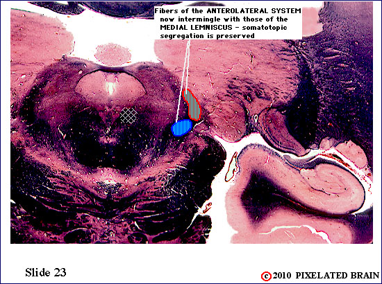
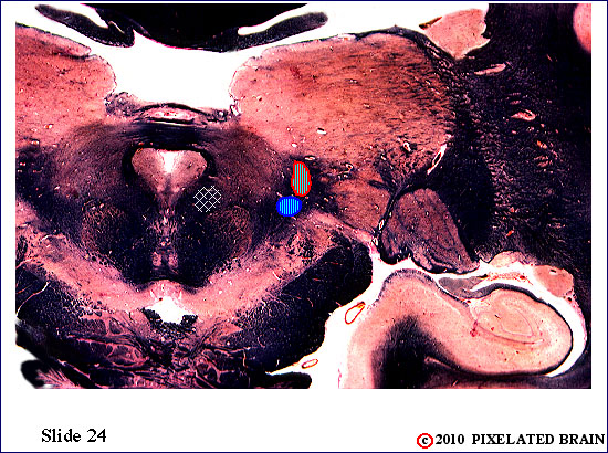
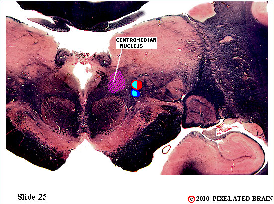
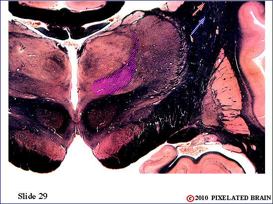
| At this level fibers of the anterolateral pathway intermingle with those of the medial lemniscus. Somatotopic order is preserved as these pathways approach their termination in VPL. The central tegmental tract has now terminated in the centromedian nucleus ( the most prominent of the intralaminar nuclei). | ||
 |
 |
|
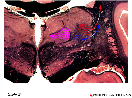
| The anterolateral pathway has now terminated in the VPL nucleus. | ||
 |
 |
|
| The anterolateral pathway has terminated in the VPL nucleus. | ||
 |
 |
|
 |
 |
|
29 |
||