|
MODULE 3 - SECTION 4 - THE THIRD ORDER NEURONS of the DCML
|
1
 |
 |
|
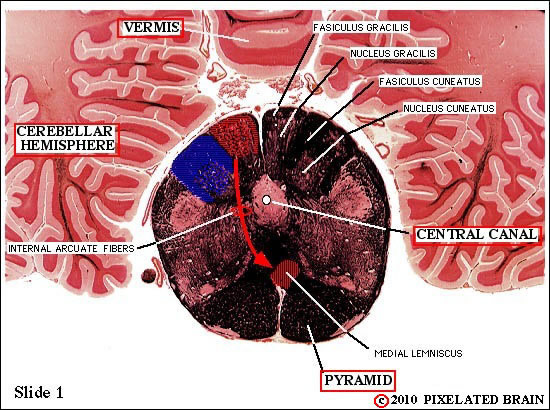
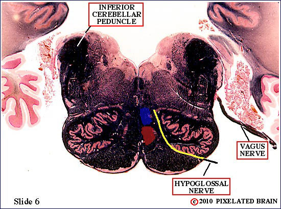
This slide passes through the caudal medulla. Second order neurons, within the nucleus gracilis give rise to fibers which decussate and begin to pile up ventrally as the first component of the medial lemniscus.
We will remind you repeatedly of the great clinical importance of knowing the level at which motor and sensory pathways cross the midline. For the DC-ML pathway the decussation occurs here, at the level of the caudal medulla. |
|
2
 |
 |
|
| |
|
3
 |
 |
|
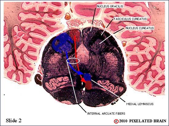
Formation of the medial lemniscus is now almost complete. It seems to be squeezed into a vertical band as it tries to get by the two inferior olives. Note that the central canal of the spinal cord is now opening up to form the fourth ventricle, roofed by choroid plexus.
Recognize that at this level, sensory information from the lower part of the body (red) travels in the ventral part of the medial lemniscus, and information from the upper part of the body travels in the more dorsal (blue) part of the structure.
|
|
4
 |
 |
|
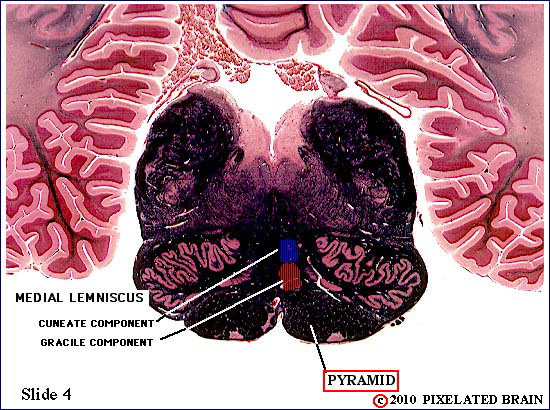
| Formation of the medial lemniscus is complete. |
|
5
 |
 |
|
| Little change here. This is a classical section through the mid-medulla. |
|
6
 |
 |
|
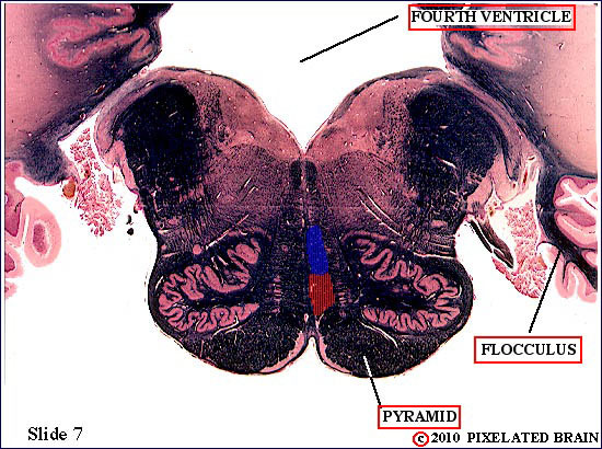
| Still the mid-medulla, and little change. Note the openings, laterally, which allow CSF to escape from the fourth ventricle into the subarachnoid space. Note that an exiting cranial nerve passes right by the medial lemniscus; why might that relationship be important? |
|
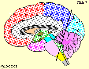
7
 |
 |
|
| |
|
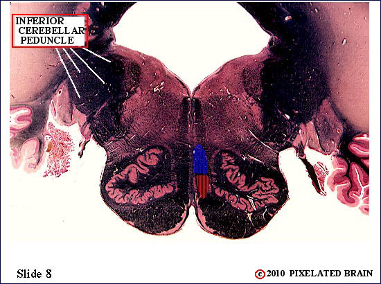
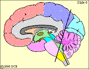
8
 |
 |
|
| Little change. The medial lemniscus is still "pressed against the midline" and held in a vertical position by the inferior olive. |
|
9
 |
 |
|
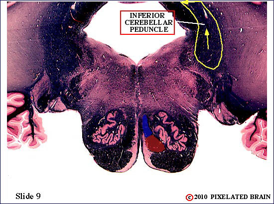
We are approaching the border between the medulla and the pons. The fourth ventricle is getting smaller. We have almost reached the rostral pole if the inferior olive. As this nucleus gets smaller, the lower part of the medial lemniscus will begin to "slide" laterally.
|
|
10
 |
 |
|
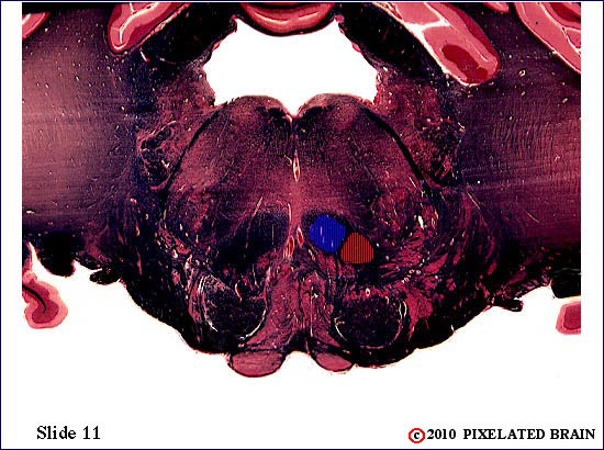
| We are approaching the border between the medulla and the pons. The fourth ventricle is getting smaller. The inferior olive is almost gone, and the lateral drift of the medial lemniscus is beginning. The component contributed by the Nucleus Gracilis (red = lower body) has been in the most ventral part of the lemniscus till now; as the lemniscus assumes a horizontal orientation, the red fibers will lie most laterally. Note the close relationship of the medial lemniscus to the pyramid. |
|
11
 |
 |
|
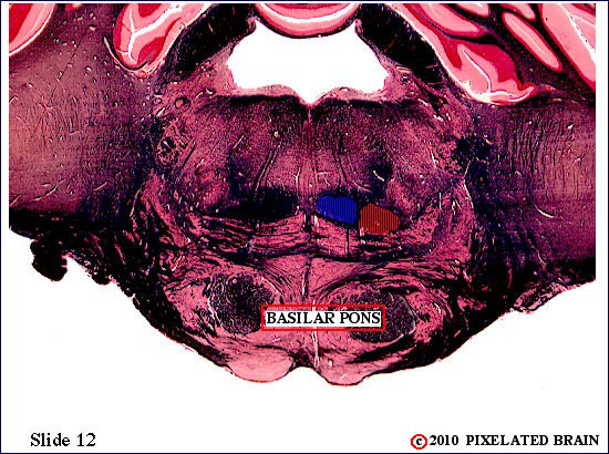
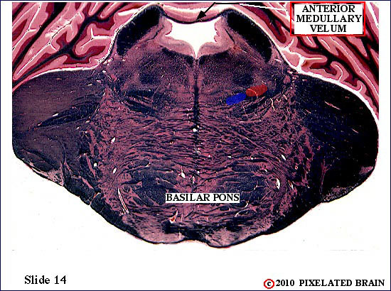
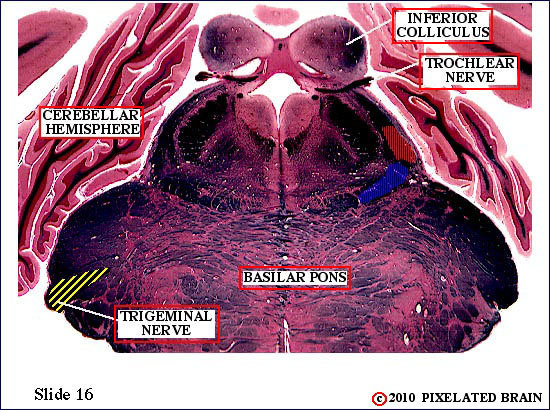
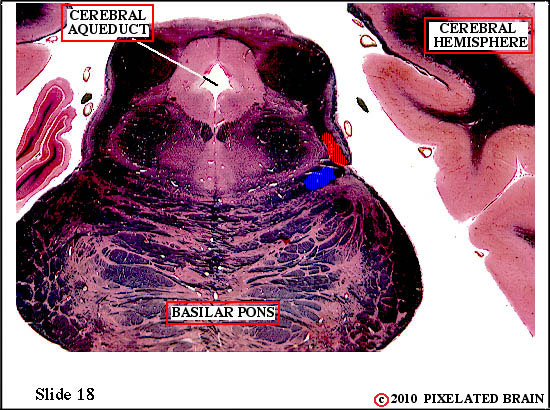
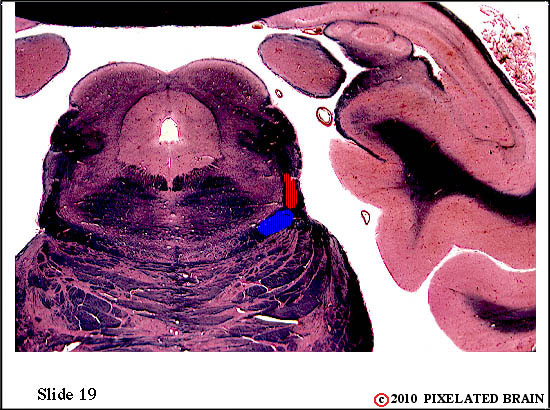
We now pass from the medulla to the pons. Note that the ventral part of the brainstem now looks a bit different.. That's because of the appearance of a "bump" on the ventral aspect of the pons, called the "basilar pons". You can see it in the sagittal view at the left. The medial lemniscus ascends through the more dorsal partof the pons in a regenion referred to as the tegementum.
Now that the inferior olive is gone, the medial lemniscus begins to move laterally; as it does so the gracile (= red = lower body) component lies lateral to the cuneate component
|
|
12
 |
 |
|
| The medial lemniscus has completed its 90 degree rotation into a horizontal position. The basilar pons is getting bigger. |
|
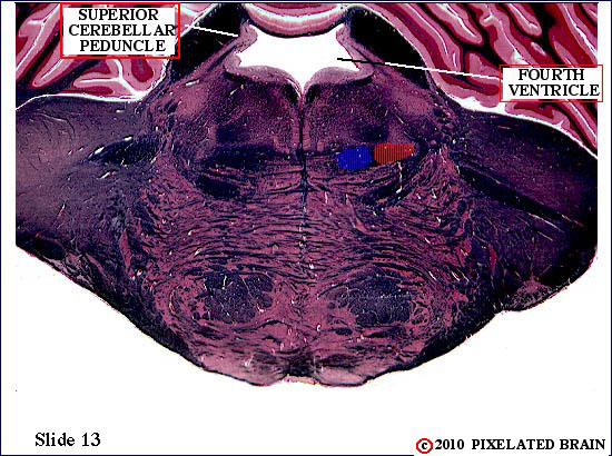
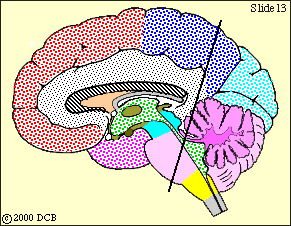
13
 |
 |
|
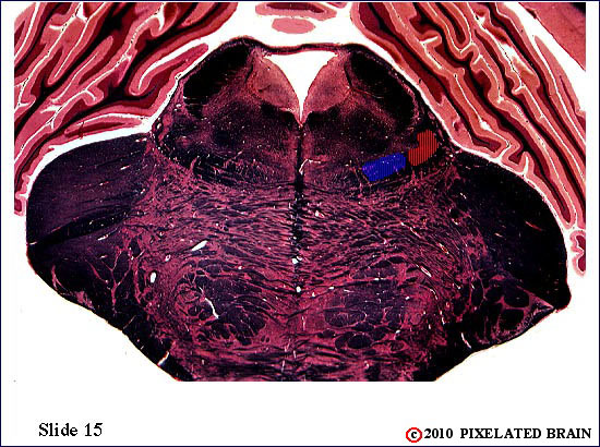
| Little change. The fourth ventricle is getting smaller and will soon be transformed into the cerebral aqueduct. |
|
14
 |
 |
|
The medial lemniscus is still horizontal and is moving away form the midline. Somatotopic organization is preserved.
The sagittal view at the left tells you that this section still passes through the pons (purple), but also that the more dorsal part of the section is getting very close to the midbrain (blue). One indication of this is that the fourth ventricle is now getting small and will soon become the cerebral aqueduct.
To identify adjacent structures, click on one of the atlas views.
|
|
15
 |
 |
|
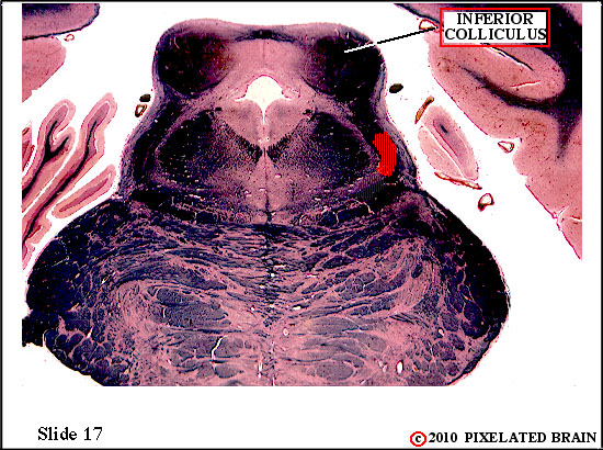
| The medial lemniscus has reachd the lateral edge of the brainstem. |
|
16
 |
 |
|
| The dorsal part of this section passes through what division of the brainstem? How can that be, since the ventral part of the section clearly passes through the pons? |
|
17
 |
 |
|
| The medial lemniscus begins to rotate a bit more. |
|
18
 |
 |
|
| |
|
19
 |
 |
|
| Little change. |
|
20
 |
 |
|
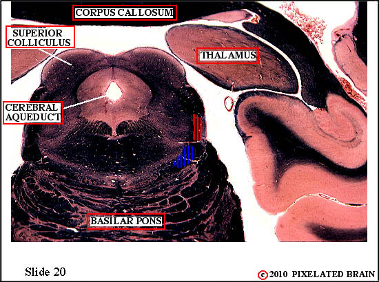
| The medial lemniscus is now passing through the tegmentum of the midbrain and approaching the thalamus. Give two reasons why you know the dorsal part of this section of the brainstem goes through the midbrain. |
|
21
 |
 |
|
| The thalamus is getting larger and has extended caudally, to "overhang" the midbrain. |
|
22
 |
 |
|
| |
|
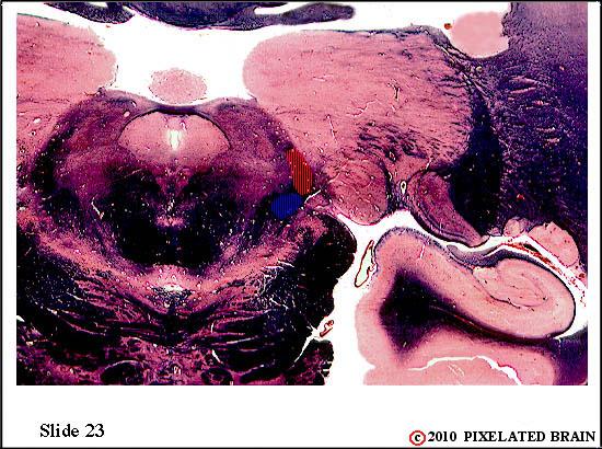
23
 |
 |
|
| |
|
24
 |
 |
|
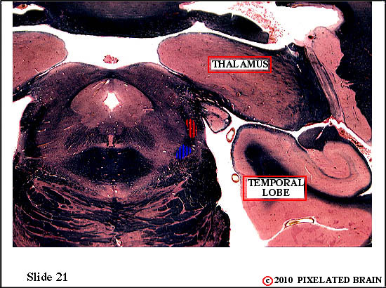
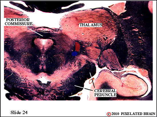
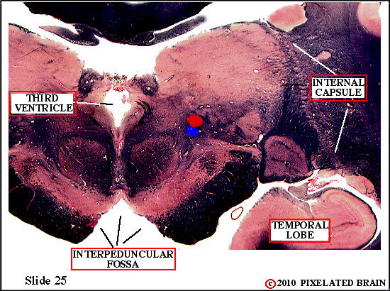
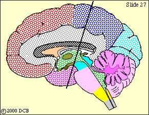
Well, we call this one of our "trifecta" slides. The sagittal view may help you understand that the ventral part of the slide passes through the basilar pons (mostly clipped off in this view), the mid portion of the slide passes through the tegmentum of the midbrain, and the most dorsal part of the slide passes through the thalamus. This is spelled outmore specifically in the lo-mag atlas view of Atlas 3.
Here, the medial lemniscus runs in the tegmentum of the midbrain. There are some interesting structures nearby: you are not "responsible" for them at this point, but click on Atlas 1 just for fun.
|
|
25
 |
 |
|
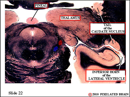
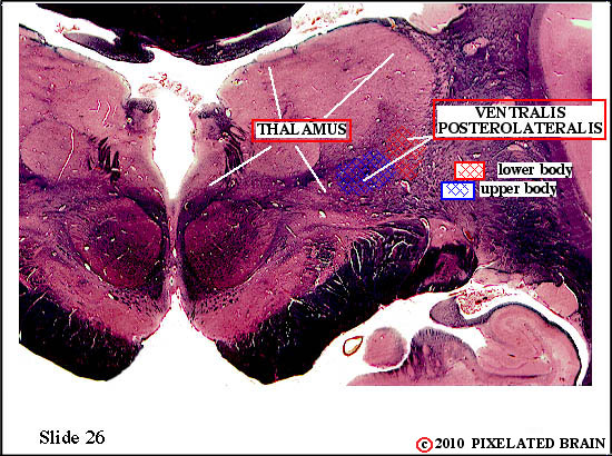
| The axons of the second order neurons in the medial lemniscus, that we have been tracing up from the medulla, are about to enter their thalamic relay nucleus. |
|
26
 |
 |
|
| It's happened! The thalamic relay nucleus for the medial lemniscus is the nucleus ventralis posterolateralis (VPL). The cell bodies of the third order neurons of the pathway are found here. |
|
27
 |
 |
|
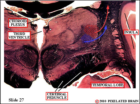
Axons of the ascending medial lemniscus pathway (second order neurons) have now terminated by synapsing on the cell bodies ot third order neurons in the nucleus ventralis posterolateralis (VPL) Third order neurons in the lateral part of this nucleus (red) relay information from the lower part of the body; neurons in the more medial part of the nucleus (blue) relay information from the upper part of the body. Both send axons laterally to enter the internal capsule.
Now would be a good time to take another look at FIGURE 3-2 and remind yourself of just where we are. |
|
28
 |
 |
|
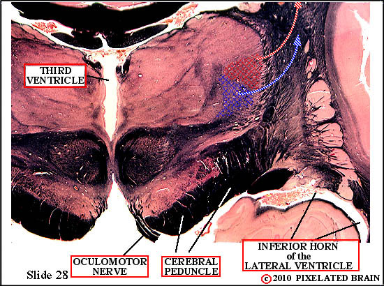
| Third order sensory neurons in VPM send axons laterally, into the posterior part of the posterior limb of the internal capsule. They will ascend to terminate in the primary sensory cortex. |
|
29
 |
 |
|
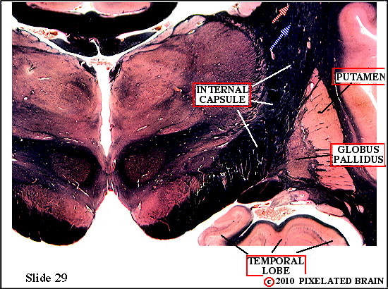
This slide passes rostral to VPL. Axons of third order neurons of the DC-ML pathway are now ascending to the cortex within the posterior limb of the internal capsule.
|
29 |
30
|
SITE MAP MOD 3 HOME PIXBRAIN HOME I WANT TO COMMENT
NEXT
|
 |
|
|