FIGURE 2-29
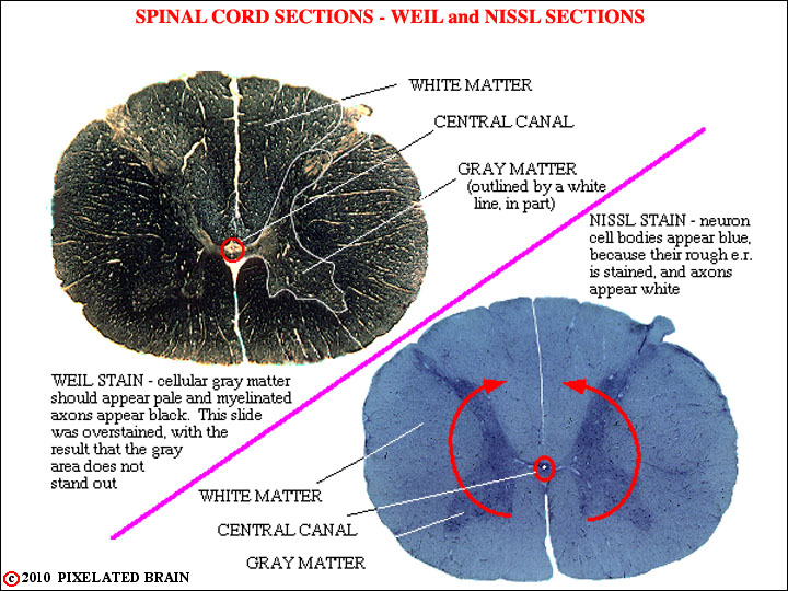
The remaining views of this section deal with the histological appearance of the spinal cord. This first view shows the general appearance of the spinal cord. The red arrows are there to remind you that it was the folding of the neural plate that gave rise to the neural tube, of which the spinal cord is a part. The cavity within the tube is quite small at spinal cord levels (the central canal), but greatly expands within some parts of the brain, as you already have discovered. At any level, the cord may be divided into a central, butterfly-shaped region - the gray matter - which contains many cells, and a peripheral zone of white matter, consisting mainly of myelinated and unmyelinated axons.