FIGURE 2-34
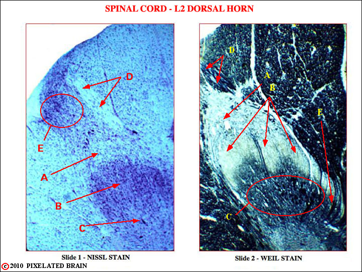
Slide 1 - L2, dorsal horn. Nissl stain. Cell bodies appear pink (because their rough e. r. is stained) and axons ("white matter") appear white. See Fig. 33 for orientation of section.
A. Posterior marginal nucleus (lamina I)
B. Substantia gelatinosa (laminae II)
C. Nucleus proprius (laminae III, IV)
D. Myelinated dorsal root fibers on their way to terminate in deeper spinal cord lam
E. Dorsolateral fasciculus (of Lissauer). Unmyelinated dorsal root fibers enter in this more lateral position and may run up or down the cord for a few segments before turning medially to terminate in laminae I, II, III.
Slide 2 - L2 dorsal horn. Weil stain. Cellular gray matter appears pale, while myelinated axons stain black. Almost same level as previous slide.
A. Posterior marginal nucleus (lamina I).
B. Substantia gelatinosa (laminae II)
C. Nucleus proprius (laminae III, IV)
D. Myelinated dorsal root fibers on their way to terminate in deeper spinal cord laminae
E. Note myelinated dorsal root axons turning laterally to terminate directly in the deeper lamine.