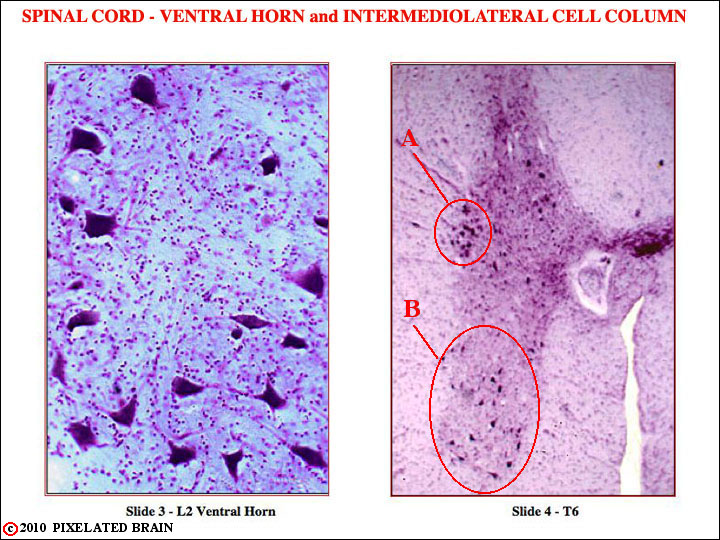
FIGURE 2-35
Slide 3 - L2 ventral horn, higher magnification. Nissl stain. Note the large cell bodies of motor neurons in what must be lamina IX. See Fig. 33 for orientation of section.
Slide 4. T6. low magnification, Nissl stain. See Fig. 33 for orientation of section.
A. Intermediolateral cell column. The cell bodies we see are those of sympathetic preganglionic neurons.
B. Motor neuron nuclei in ventral horn. Note that at this thoracic level, where the motor innervation is of trunk muscles only (no extremity), the anterior horn is rather narrow. Compare with the anterior horn at the level of C6, shown in Fig. 2-37.