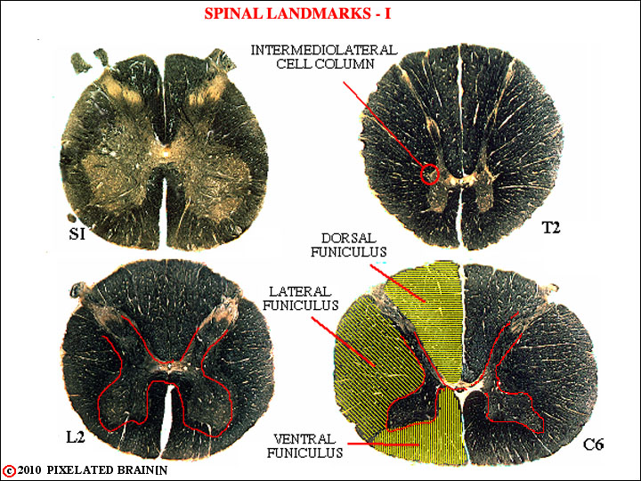
FIGURE 2-30
This view shows several features of spinal cord anatomy that are helpful in placing the level of any given section.
1) The gray matter enlarges at the level of the lumbosacral plexus (S1 - L2) and at the level of the brachial plexus (C5-C8)). It is relatively sparse at thoracic levels.
2) White matter occupies an increasing fraction of the total area as the cord approaches the brainstem.
3) The intermediolateral cell column is present only from T1 to L2 or L3.
The pathways which connect the spinal cord with the brain ascend and descend within the white matter. This peripheral region is divided by the dorsal and ventral horns of gray matter into three funiculi - the dorsal funiculus, the lateral funiculus and the ventral funiculus. At upper thoracic and all cervical levels the dorsal funiculus is further subdivided, as shown in the next view Figure 2-31.