- PIXBRAIN HOME _ _ MOD 6 HOME _ _ I WANT TO -
caudal___SLIDE L2 ___rostral
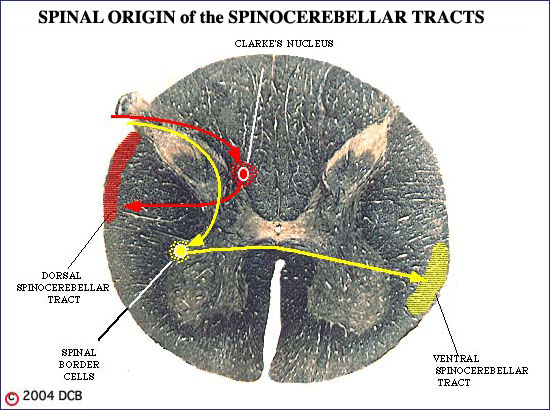
THE DORSAL SPINOCEREBELLAR TRACT (DSCT)
First order neurons associated with muscle spindles, Golgi tendon organs and touch and pressure receptors in the leg send fibers into the cord to terminate in Clarke's Nucleus. Since the nucleus is present only from Ti toL3, afferents from more caudal segments have to ascend in the cord to reach it. Second order neurons within the nucleus send their axons laterally to ascend near the surface of the cord in the posterior part of the lateral funiculus. This pathway sends information about position and movement of the leg to the intermediate zone of the ipsilateral hemisphere.
THE VENTRAL SPINOCEREBELLAR TRACT (VSCT)
Cells on the lateral surface of the anterior horn of the lumbar cord give rise to the ventral spinocerebellar tract. Because of their position, these are called spinal border cells. These second order neurons receive the same inputs as described above for the DSCT, but in addition they receive inputs from a wide variety of cutaneous receptors and from descending motor pathways - primarily, the pyramidal tract. Thus they are "aware" not only of leg position and movement, but also of the motor "commands" that have initiated the movement. The ascending route taken by VSCT is also remarkable, as shown in Figure 6-16. The axons cross the midline to ascend on the contralateral side of the cord and brainstem, then cross back and enter the ipsilateral cerebellar hemisphere traveling with the superior cerebellar peduncle.