REVIEW #2
In this review, we look at 16 labeled slides that are also a part of Review #1. In this case, questions are asked about several structures on each slide. Question views alternate with ones providing answers.

QUESTION 1
2Name three places where the fibers in this pathway terminate.
What loss would result if the pathway were cut at the point where the pointer touches it?
3) Important vessels enter the brain in this region; give their name(s) and give one structure that they supply.
Give two symptoms that result when this structure is damaged.
ANSWER 1
2) Name three places where the fibers in this pathway terminate. The amygdala, the periamygdaloid cortex and the contralateral olfactory bulb.
What loss would result if the pathway were cut at the point where the pointer touches it? The subject would be unable to detect odors inhaled through the ipsilateral nostril. Testing might be tricky, however, because of the way in which air circulates at the back of the nasal cavity. Also, strong odors are often "sensed" by the taste receptors of the tongue.
3) Important vessels enter the brain in this region; give their name(s) and give one structure that they supply. Medial and lateral striates or lenticulostriates. They supply the caudate, putamen, globus pallidus and much of the internal capsule
11) Identify this small, dark purple structure with wiggly edges. The flocculus.
Give two symptoms that result when this structure is damaged. Nystagmus, vertigo, difficulty with balance.

QUESTION 2
4) Destruction of the gyrus lying just above this sulcus on the right side of the brain (right hemisphere) would result in what sensory loss?
9) Identify this cranial nerve.
10) What (output) pathway originates in this structure,and where does this pathway terminate?
ANSWER 2
4) Destruction of the gyrus lying just above this sulcus on the right side of the brain (right hemisphere) would result in what sensory loss? A left homonymous inferior quadrantanopia.
9) Identify this cranial nerve. The oculomotor nerve.
10) What (output) pathway originates in this structure,and where does this pathway terminate? The mammilothalamic tract. It will terminate in the anterior nucleus of the thalamus.

QUESTION 3
1) Name this space.
Describe the path of CSF as it leaves this space, and, ultimately, returns to the venous system.
8) What sensory pathway passes through this structure?
What loss would result if this structure were badly damaged on the left side of the brainstem?
14) Identify this thalamic nucleus.
What would happen to the pupillary light reflex if this nucleus were damaged on the left side of the brain?
ANSWER 3
1) Name this space. The anterior horn of the lateral ventricle.
Describe the path of CSF as it leaves this space, and, ultimately, returns to the venous system. Interventricular foramen (of Monro) -- third ventricle -- cerebral aqueduct -- fourth ventricle -- foramen of Lushka and Magendie -- subarachnoid space -- arachnoid granulations -- superior sagittal sinus.
8) What sensory pathway passes through this structure? The auditory pathway.
What loss would result if this structure were badly damaged on the left side of the brainstem? Little if any, because auditory information from each ear ascends on both sides of the brainstem.
14) Identify this thalamic nucleus. The lateral geniculate nucleus.
What would happen to the pupillary light reflex if this nucleus were damaged on the left side of the brain? It would be normal.

QUESTION 4
4) Identify this surface "bump".
What nucleus lies just deep to this landmark?
7) This cranial nerve is exiting through the middle cerebellar peduncle; what motor loss would result if it were cut on the left side of the brainstem?
What effect would the same cut have on: a) the pupillary light reflex and b) the corneal reflex?
9) What part of the motor cortex (from a somatotopic point of view) is normally supplied by this blood vessel?
Is this vessel a part of the anterior or posterior systems?
ANSWER 4
4) Identify this surface "bump". The uncus.
What nucleus lies just deep to this landmark? The amygdala.
7) This cranial nerve is exiting through the middle cerebellar peduncle; what motor loss would result if it were cut on the left side of the brainstem? The muscles of mastication (and a few others) on the left side of the head would fail to contract (lower motor neuron paralysis).
What effect would the same cut have on: a) the pupillary light reflex and b) the corneal reflex? No effect on the pupillary light reflex. The corneal reflex would be abnormal: touching the left cornea (with a wisp if cotton) would result in no eye closure; touching the right cornea would result in both eyes closing.
9) What part of the motor cortex (from a somatotopic point of view) is normally supplied by this blood vessel? The leg area.
Is this vessel a part of the anterior or posterior systems? The anterior circulation.

QUESTION 5
2) Name this nucleus.
Neurons here send their axons to terminate in what nucleus? (include the word ipsi/contralateral in your answer)
5) Name this pathway.
In what structure and "zone" will these axons terminate?
10) Name this structure.
Neurons here send their axons to the cerebellum. Through which peduncle do they travel and with what cells in the cerebellar cortex do they make synaptic contact? (include the word ipsi/contralateral in your answer)
ANSWER 5
2) Name this nucleus. The nucleus gracilis.
Neurons here send their axons to terminate in what nucleus? (include the word ipsi/contralateral in your answer) The contralateral nucleus ventralis posterolateralis of the thalamus.
5) Name this pathway. The dorsal spinocerebellar tract.
In what structure and "zone" will these axons terminate? The intermediate zone and vermis of the cerebellar cortex.
10) Name this structure. The inferior olive.
Neurons here send their axons to the cerebellum. Through which peduncle do they travel and with what cells in the cerebellar cortex do they make synaptic contact? (include the word ipsi/contralateral in your answer) The axons cross the midline and pass through the contralateral inferior cerebellar peduncle to enter the cerebellum. The axons terminate as climbing fibers, making synaptic contact with purkinje cells.

QUESTION 6
3) Identify the nucleus found in this region. Also give the letters often used (GSA, etc.) to describe its function.
Describe, in words, the function of the neurons in this nucleus.
6) How would you test this nerve?
Describe the nature of the corticobulbar input to this nucleus?
10) What motor nucleus is found in this region?
Neurons here contribute axons to which cranial nerve(s)?
ANSWER 6
3) Identify the nucleus found in this region. Also give the letters often used (GSA, etc.) to describe its function. The dorsal motor nucleus of the vagus. (GVE).
Describe, in words, the function of the neurons in this nucleus. These are are parasympathetic preganglionic neurons.
6) How would you test this nerve? This is the hypoglossal nerve. Ask the subject to stick out her tongue. If nerve function is impaired, the tongue will deviate to the impaired side. Atrophy and fasciculations indicate a lower motor neuron problem. Lack of these signs suggests damage to the corticobulbar pathway descending from the contralateral hemisphere.
Describe the nature of the corticobulbar input to this nucleus? The corticobulbar pathway is bilateral, but more crossed than uncrossed.
10) What motor nucleus is found in this region? The nucleus ambiguus.
Neurons here contribute axons to which cranial nerve(s)? The glossopharyngeal and vagus.

QUESTION 7
1) Identify this nucleus. Also give the letters used to describe its function.
A lesion here would also destroy the fibers of a cranial nerve (labeled #2) that loops around the nucleus. What motor loss would result because of damage to this second structure?
3) What somatic sensory pathway ascends in this area?
Where do the axons in this pathway have their cell bodies? (include the word ipsi/contralateral in your answer).
What loss would result if the axons marked by the yellow and green dots were cut?
ANSWER 7
1) Identify this nucleus. Also give the letters used to describe its function. The abducens nucleus (SE)
A lesion here would also destroy the fibers of a cranial nerve (labeled #2) that loops around the nucleus. What motor loss would result because of damage to this second structure? That's the facial nerve. There would be a lower motor neuron type paralysis of all the muscles of facial expression (plus a few others).
3) What somatic sensory pathway ascends in this area? The anterolateral pathway.
Where do the axons in this pathway have their cell bodies? (include the word ipsi/contralateral in your answer). In the contralateral dorsal horn.
What loss would result if the axons marked by the yellow and green dots were cut? These are the axons of cells in the nucleus cuneatus. They convey the senses of touch, vibration, position from the upper part of the body on the contralateral side.

QUESTION 8
1) Identify this compact bundle of fibers.
Where do these axons have their cell bodies?
2) What ascending somatic sensory pathway is found in this region?
In what nucleus will the axons of this pathway terminate?
3) Identify this motor nucleus and give the letters used to describe its function.
What other motor nuclei lie in the same functional cell column?
ANSWER 8
1) Identify this compact bundle of fibers. It is the mesencephalic root of the trigeminal nerve.
Where do these axons have their cell bodies? In the nucleus of the mesencephalic root.
2) What ascending somatic sensory pathway is found in this region? The ventral secondary trigeminal tract.
In what nucleus will the axons of this pathway terminate? The nucleus ventralis posteromedialis (for touch, sharp pain, and temperature) and the intralaminar nucleus (for dull aching pain ).
3) Identify this motor nucleus and give the letters used to describe its function. The motor nucleus of the trigeminal nerve. (SVE)
What other motor nuclei lie in the same functional cell column? The facial nucleus, the nucleus ambiguus and the accessory nucleus.

QUESTION 9
2) Identify this pathway.
Some of the fibers within this bundle are part of a somatic sensory pathway. Where will they terminate and what sorts of sensations do they convey?
4) Give the input and output pathways for this structure.
What word is used to describe how the neurons in this structure are organized.
5) Describe how this cranial nerve exits from the skull.
What muscle(s) does it innervate?
ANSWER 9
2) Identify this pathway. The central tegmental tract.
Some of the fibers within this bundle are part of a somatic sensory pathway. Where will they terminate and what sorts of sensations do they convey? The pathway is the so-called indirect spinothalamic one. The fibers will terminate in the intralaminar nuclei (= centromedian) and they convey a sense of dull, aching pain.
4) Give the input and output pathways for this structure. This is the inferior colliculus. The input pathway is the lateral lemniscus and the output pathway is the brachium of the inferior colliculus.
What word is used to describe how the neurons in this structure are organized. Tonotopic
5) Describe how this cranial nerve exits from the skull. This is the trochlear nerve. It departs from the dorsal aspect of the brainstem, wraps around the brainstem, passes through the cavernous sinus, and the through the superior orbital fissure to enter the orbit.
What muscle(s) does it innervate? The superior oblique muscle.

QUESTION 10
4) Where do these axons have their cell bodies?
Describe what would happen to the pupillary light reflex if this pathway were cut on the right side of the brain. Also, what would happen to the corneal reflex?
5) Where will this pathway terminate? Where did it originate?
It's not very obvious - so what's the best way to find it?
8) Identify this "C shaped" structure.
ANSWER 10
4) Where do these axons have their cell bodies? In the retina.
Describe what would happen to the pupillary light reflex if this pathway were cut on the right side of the brain. Also, what would happen to the corneal reflex? Because of the chiasm and the posterior commissure, light shined in either eye will still activate both Edinger-Westphal nuclei - so the pupillary light reflex will be normal. Obviously the corneal reflex will be normal as well.
5) Where will this pathway terminate? Where did it originate? This is the stria terminalis. It originates in the amygdala and terminates in the septal area and the hypothalamus.
It's not very obvious - so what's the best way to find it? It (almost) always lies just medial to the tail of the caudate nucleus.
8) Identify this "C shaped" structure. This is the dentate nucleus

QUESTION 11
4) In what "division" of the brain is this nucleus found? What pathway provides the major input to the nucleus?
6) What pathway descends in this region?
Damage to this pathway is associated with a classical group of symptoms. Give three of them?
9) Identify this nucleus.
Describe the clinical picture that results when this nucleus is damaged on the left side of the brainstem.
ANSWER 11
4) In what "division" of the brain is this nucleus found? What pathway provides the major input to the nucleus? This is the lateral geniculate nucleus nucleus. It is in the diencephalon and the optic tract provides the major input.
6) What pathway descends in this region? The corticospinal, or pyramidal, tract.
Damage to this pathway is associated with a classical group of symptoms. Give three of them? Hyperactive deep tendon reflexes, increased resistance to passive stretch, clonus, "positive" Babinski response (the plantar response would be extensor)
9) Identify this nucleus. The oculomotor nucleus.
Describe the clinical picture that results when this nucleus is damaged on the left side of the brainstem. The lid will be closed. If you pull it up, the eye will be seen to be abducted and depressed. (While you won't be able to see it, in will be rotated medially = intorsion). The pupil will be dilated and fail to react to light.

QUESTION 12
1) Identify this nucleus.
This nucleus is one of many that could be considered a part of the limbic system. Why is that?
2) Identify this nucleus.
Of which cerebellar "loop" or circuit is this nucleus a part?
10) Damage to this nucleus gives rise to what movement disorder?
Will the abnormal movements be ipsilateral or contralateral to the damaged nucleus?
ANSWER 12
1) Identify this nucleus. The medialis dorsalis.
This nucleus is one of many that could be considered a part of the limbic system. Why is that? Because it receives a significant input from the amygdala by way of the ventral amygdalofugal path.
2) Identify this nucleus. Ventralis lateralis
Of which cerebellar "loop" or circuit is this nucleus a part? The "planning" loop.
10) Damage to this nucleus gives rise to what movement disorder? Hemiballismus.
Will the abnormal movements be ipsilateral or contralateral to the damaged nucleus? Contralateral.

QUESTION 13
6) Identify this nucleus.
Give 3 abnormal findings (symptoms) associated with destruction of this nucleus in experimental animals.
8) Identify this general region of the brain.
Give three sorts of problems that might result following damage to this region.
11) Identify this tract.
It is one component of a famous neural "circuit". What is the name of the circuit?
ANSWER 13
6) Identify this nucleus. The amygdala
Give 3 abnormal findings (symptoms) associated with destruction of this nucleus in experimental animals. Descriptions of this "Kluver-Bucy" syndrome vary slightly but here is one list. These animals: 1)are fearless and placid, lacking in emotional reactions 2) do not react to threatening situations 3) display indiscriminate hypersexual behavior 4) are inordinately attentive to all stimuli = ceaselessly curious 5) examine objects with their mouth rather than the eyes; eat food normally avoided 6) seem to recognize nothing (= visual agnosia)
8) Identify this general region of the brain. Hypothalamus.
Give three sorts of problems that might result following damage to this region. Problems with control of: 1) temperature, 2) food intake, 3) autonomic functions, 4) pituitary function.
11) Identify this tract. The mammillothalamic tract.
It is one component of a famous neural "circuit". What is the name of the circuit? The Papez circuit.

QUESTION 14
1) Identify this gyrus.
6) Name one place where the fibers in this "peduncle" will terminate.
8) Identify this structure.
10) Identify this pathway.
11) What is this space, and how does CSF exit from it?
ANSWER 14
1) Identify this gyrus. The cingulate gyrus
6) Name one place where the fibers in this "peduncle" will terminate. The red nucleus; the nucleus ventralis
8) Identify this structure. The inferior olive.
10) Identify this pathway. The fornix.
11) What is this space, and how does CSF exit from it? The anterior horn of the lateral ventricle. CSF exits by passing through the interventricular foramen into the third ventricle.

QUESTION 15
1) Identify this structure.
Where did most of these fibers originate and where will most of them terminate?
5) Identify this structure.
Where does the major input to this structure come from?
8) Identify this structure.
10) Identify this fissure.
ANSWER 15
1) Identify this structure. The fornix.
Where did most of these fibers originate and where will most of them terminate? Cells in the subiculum contribute most of the axons present in the fornix. The most obvious group of fibers terminate in the mammillary body. But fibers also terminate elsewhere in the thalamus, hypothalamus and septum.
5) Identify this structure. The putamen.
Where does the major input to this structure come from? The cerebral cortex.
8) Identify this structure. The anterior commissure.
10) Identify this fissure. The lateral fissure.
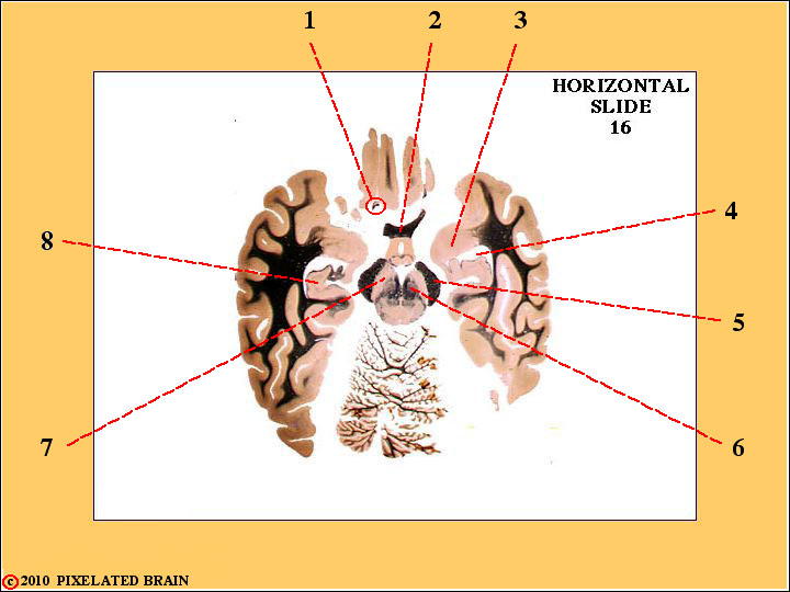
QUESTION 16
1) Identify this structure.
2) Identify this structure.
3) Identify this nucleus
4) Identify this space.
8) Identify this structure.
ANSWER 16
1) Identify this structure. The olfactory tract.
2) Identify this structure. The optic chiasm.
3) Identify this nucleus. The amygdala.
4) Identify this space. The inferior horn of the lateral ventricle.
8) Identify this structure. The hippocampus.
This is the last slide of Review 2.